Download
1 / 30
340 likes | 2.17k Views
Normal Radiographic Anatomy of the Equine metacarpus/metatarsus, fetlock, and distal extremity. First Year Radiology. Dr Virginie De Busscher. Metacarpus(MC)/Metatarsus(MT). MC and MT are similar Large nutrient foramen in MC/MT III “splint bones”: 2 nd and 4 th MC/MT

E N D
Normal Radiographic Anatomy of the Equinemetacarpus/metatarsus, fetlock, and distal extremity First Year Radiology Dr Virginie De Busscher
Metacarpus(MC)/Metatarsus(MT) • MC and MT are similar • Large nutrient foramen in MC/MT III • “splint bones”: 2nd and 4th MC/MT • Dorsopalmar(plantar) view: marker placed laterally • Lateral views: marker is dorsal • 4 views • Multiple oblique views if fracture suspected
Metacarpus/Metatarsus Dorsopalmar(plantar) view A: Second MC/MT B: Fourth MC/MT C: Third MC/MT D: Medial proximal sesamoid bone E: Lateral proximal sesamoid bone 1: Nutrient foramen 2: MC/MT phalangeal joint L From Ulg (Belgium) From KSUCVM
Metacarpus/Metatarsus Lateromedial view A: Second MC/MT B: Fourth MC/MT C: Third MC/MT D: Medial proximal sesamoid bone E: Lateral proximal sesamoid bone 1: Nutrient foramen 2: MC/MT phalangeal joint
Dorsal Lateral Medial Palmar/Pl Metacarpus/Metatarsus DLPM oblique view DLPMo = DorsoLateral-PalmaroMedial oblique L B: Fourth MC/MT C: Third MC/MT D: Medial proximal sesamoid bone E: Lateral proximal sesamoid bone
Metacarpus/Metatarsus DMPL oblique view DMPLo = Dorsomedial-PalmaroLateral oblique Dorsal L Lateral Medial Palmar/Pl A: Second MC/MT C: Third MC/MT D: Medial proximal sesamoid bone E: Lateral proximal sesamoid bone
Metacarpophalangeal/Metatarsophalangeal Articulation Fetlock • Dorsopalmar(plantar) views: • impossible to distinguish med/lat without marker • Marker has to be lateral • Lateral views: marker is dorsal • Few differences between forelimb and hindlimb • 4 views + lateromedial view in flexion if needed
Metacarpophalangeal/Metatarsophalangeal Articulation Lateromedial view A: Third MC/MT bone B: Proximal phalanx (P1) 1: Sagittal ridge 3: Palmar process of proximal phalanx 4: Condyles of third metacarpal bone 5: Proximal sesamoid bone
Metacarpophalangeal/Metatarsophalangeal Articulation Dorsopalmar(plantar) view R A: Third MC/MT bone B: Proximal phalanx (P1) C: Medial proximal sesamoid bone D: Lateral proximal sesamoid bone E: Metacarpo(tarso)-phalangeal joint F: Middle phalanx (P2) G: Third phalanx (P3) H: Proximal interphalangeal joint I: Distal interphalangeal joint
Metacarpophalangeal/Metatarsophalangeal Articulation Dorsopalmar(plantar) view R To prevent superimposition of the proximal sesamoid bones over the joint, the beam should be descending Dorsoproximal-Palmarodistal oblique view
Metacarpophalangeal/Metatarsophalangeal Articulation Dorsopalmar(plantar) view R A: Third MC/MT bone B: Proximal phalanx C: Medial proximal sesamoid bone D: Lateral proximal sesamoid bone E: Metacarpo(tarso)-phalangeal joint J: Depression for medial collateral ligament attachment 1: Sagittal ridge Sagittal ridge at MC/MT distal aspect
Metacarpophalangeal/Metatarsophalangeal Articulation: Dorsopalmar view R MC/MT distal aspect P1 dorsal P1 proximal P1 palmar/plantar P2 dorsal P2 palmar/plantar
Dorsal Lateral Medial Palmar/Pl DLPMo Metacarpophalangeal/Metatarsophalangeal Articulation DLPM oblique view A: Third MC/MT bone B: Proximal phalanx C: Medial proximal sesamoid bone D: Lateral proximal sesamoid bone E: Metacarpo(tarso)-phalangeal joint 1: Sagittal ridge 3: Palmar process of proximal phalanx/Lateral palmar tubercle (eminence) 6: Medial condyle of third MC/MT bone R Beam can be slightly descending (proximo-distal)
Dorsal Lateral Medial Palmar/Pl Metacarpophalangeal/Metatarsophalangeal Articulation DMPL oblique view A: Third MC/MT bone B: Proximal phalanx C: Medial proximal sesamoid bone D: Lateral proximal sesamoid bone E: Metacarpo(tarso)-phalangeal joint 1: Sagittal ridge 2: Lateral condyle of third metacarpal/tarsal bone 3: Palmar process of proximal phalanx R DMPLo
Metacarpophalangeal/Metatarsophalangeal Articulation Forelimb Hindlimb Bulging • The MT is convex at its distal aspect DP view - Hindlimb DP view - Forelimb • The proximal sesamoid bones • are higher • The proximal sesamoid bones • are more triangular
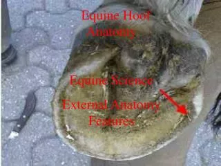
Distal Extremity Foot • No shoe, trimmed, placed on a block • For the DP views: Soap or “play-doh” (soft tissue opacity) to fill the 3 sulci of the frog avoid artifact due to air • Marker placed laterally (impossible to tell if med or lat without marker) • Use of a grid • 4 views
Distal Extremity Lateromedial view A: Middle phalanx B: Third phalanx C: Navicular bone 1: Proximal interphalangeal joint 2: Distal interphalangeal joint 3: Extensor process 4: Dorsal surface 5: Palmar process From Animalia curandi
Distal Extremity Lateromedial view Navicular bone A: Middle phalanx B: Third phalanx C: Navicular bone 1: Proximal interphalangeal joint 2: Distal interphalangeaol joint 3: Extensor process 4: Dorsal surface 5: Palmar process
Dorsal recess of the distal interphalangeal joint Extensor process Proximal margin of the navicular bone Flexor surface Proximal palmar process Articular margin Solar canal Distal palmar process Distal margin of the navicular bone Distal Extremity Lateromedial view
2 5 5 B 6 7 Distal Extremity Dorsoproximal-palmar(pl)odistal oblique view R A: Middle phalanx B: Third phalanx C: Navicular bone 1: Proximal interphalangeal joint 2: Distal interphalangeaol joint 3: Extensor process 4: Dorsal surface 5: Palmar process 6: Vascular channel 7: Solar margin Dorsoproximal-palmar(plantar)odistal oblique view
6 A C 7 B Distal Extremity Dorsopalmar(plantar) view R A: Middle phalanx B: Distal phalanx C: Navicular bone 6: Proximal interphalangeal joint 7: Distal interphalangeal joint Dorsopalmar(plantar) view
Distal Extremity Extensor process R The extensor process can have different shapes
L Proximal Distal Distal Extremity Palmar processes L
Distal Extremity Navicular bone = distal sesamoid bone A: Middle phalanx B: Distal phalanx C: Navicular bone 1: Flexor cortex 2: Proximal border 3: Articular surface 4: Distal border 5: Ridge to which impar ligament attaches
Distal Extremity Navicular bone L C: Navicular bone 1: Flexor cortex 2: Proximal border 7: Distal interphalangeal joint 7a: Palmar aspect 7b: Dorsal aspect
Articular margins Proximal and distal margins Extensor process Articular surface of third phalanx Vascular channels Crena marginis solearis Distal Extremity Navicular bone L Toe notch = Crena marginis solearis
Distal Extremity Palmaro(plantar)oproximal-palmar(plantar)odistal oblique view Navicular bone R C: Navicular bone 3: Articular surface 8: Palmar aspect of middle phalanx 9: Nutrient foramen 10: Sagittal ridge 11: Articulation between navicular bone and middle phalanx
R Dorsal DLPMo Lateral Medial Palmar/Pl Distal Extremity DLPM oblique view R
R Dorsal DMPLo Lateral Medial Palmar/Pl Distal Extremity DMPL oblique view R
Distal Extremity Hoof wall A C B B C A: Middle phalanx B: Third phalanx C: Navicular bone