Download
1 / 13
150 likes | 3.14k Views
Normal Radiographic Anatomy of the Equine Hind Limb. Dr. Pack First Year Radiology. The Equine Stifle. 2 views normally Lateral and CC Slight oblique for OCD Sedation Thick muscles Film placement. www.upei.ca/~vetrad. Equine Stifle – Lateral View. Base/Apex of patella

E N D
Normal RadiographicAnatomy of theEquine Hind Limb Dr. Pack First Year Radiology
The Equine Stifle • 2 views normally • Lateral and CC • Slight oblique for OCD • Sedation • Thick muscles • Film placement www.upei.ca/~vetrad
Equine Stifle – Lateral View • Base/Apex of patella • Medial and Lateral ridges of femoral trochlea • Extensor fossa of femur • Fibula • Tibial tuberosity www.upei.ca/~vetrad
Equine Stifle CC View • Patella • Condyles • Epicondyles • Intercondylar eminences • Fibula • Tibial tuberosity www.upei.ca/~vetrad
Things that make you go hmm … www.upei.ca/~vetrad
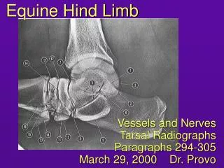
Equine Tarsus • 4 routine radiographs • Dorso-plantar • Lateral • DMPLO • DLPMO • Tarsus is very complex www.upei.ca/~vetrad
Equine Tarsal Joints • Tibiotarsal • Proximal intertarsal • Distal intertarsal • Tarsometatarsal www.upei.ca/~vetrad
Equine Tarsus DP View • Tibia • Medial malleolus • Lateral malleolus • Tibial cochlea • Talus • Medial trochlear ridge • Lateral trochlear ridge • Calcaneous www.upei.ca/~vetrad
Equine Tarsus DP View (cont) • Central tarsal bone • 2nd tarsal bone • 3rd tarsal bone • 4th tarsal bone (2 story bone) • 1st tarsal bone (occasionally seen) • 2nd, 3rd,4th metatarsal bones www.upei.ca/~vetrad
Equine Tarsus Lateral View • Sustentaculum tali • Calcaneous • Medial/lateral ridges of the trochlear talus • Tarsal bones • Proximal row • Distal row • Metatarsals www.upei.ca/~vetrad
Equine Tarsus DLPMO view • Surfaces seen? • Distal intermediate ridge of the tibia (DIRT) • Trochlear ridges • Calcaneous • Tarsal bones • Metatarsal bones www.upei.ca/~vetrad
Equine Tarsus DMPLO View • Surfaces seen? • Distal intermediate ridge of the tibia (DIRT) • Trochlear ridges • Larrys nose • Calcaneous • Tarsal bones • Metatarsal bones www.upei.ca/~vetrad
Normal RadiographicAnatomy of theEquine Hind Limb Dr. Pack www.upei.ca/~vetrad/Anatomy