Download
1 / 35
400 likes | 661 Views
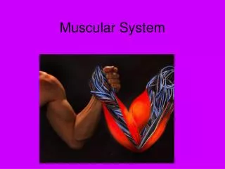
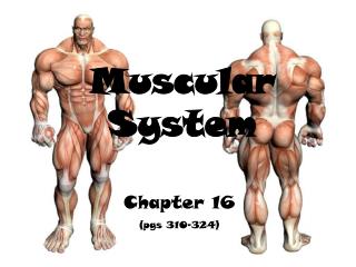
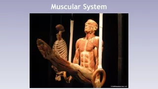
Muscular System. Skeletal – striated & voluntary. Smooth – involuntary. Types of Muscle. Cardiac - heart. Muscles and Muscle Fibers . Muscles are composed of many fibers that are arranged in bundles called FASCICLES. Fascia. Fascicle.

E N D
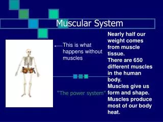
Skeletal – striated & voluntary • Smooth– involuntary Types of Muscle • Cardiac- heart
Muscles and Muscle Fibers Muscles are composed of many fibers that are arranged in bundles called FASCICLES Fascia Fascicle Individual muscles are separated by FASCIA, which also forms tendons and aponeuroses (sheet-like coverings)
Muscle Layers Muscle cell or fiber Endomysium – fascia around the cell or fiber Perimysium- fascia around a group of fibers Epimysium – fascia around the entire muscle bundle
Lets get the hierarchy straight Muscles are made from a lot of Muscle Fiber Bundles which are made of Individual Muscle Fibers (cells) which are made of Myofibrils which are rows of Sarcomeres
Muscles receive their information from the nerve cells at the Neuromuscular junction. Muscles & Nervous System
The means of contracting comes Sarcoplasmic Reticulum. It is like the smooth and rough E.R. in form and stores calcium . When the signal comes from the nerves (acetylcholine), it floods the cell with calcium which causes a chemical reaction between the thin and thick filaments.
These filaments are: – ACTIN (thin) and MYOSIN (thick) Myofibril – thick and thin filaments • These filaments overlap to form dark and light bands on the muscle fiber • A band = dArk • thick (myosin) • I band = lIght • thin (actin) • In the middle of each I band are Z lines. A sarcomere is one Z line to the other
What are the Thin and Thick filaments? Actin fibers bind to make the thin filaments Myosin fibers combine to make the thick filaments. Together they oppose each other to form the “bands” of the Sarcomere
SLIDING FILAMENT THEORY (MODEL) The theory of how muscle contracts is the sliding filament theory. The contraction of a muscle occurs as the thin filament slide past the thick filaments.
DLC the anatomy of a skeletal muscle cell from fiber to sarcomere. Page 167 will be helpful
Where does the cell get it’s energy? Fibers contain multiple mitochondria for energy ATP is made here!
Provided by ATP from cellular respiration (mitochondria) Usually only 4-6 seconds worth is stored • Aerobic respiration creates 36 ATP molecules/Glucose – must have O2 • Anaerobic Respiration creates 2 ATP and Lactic Acid – absence of O2 Energy Source
Threshold Stimulus Minimal strength required to cause a contraction Motor neuron releases enough acetylcholine to reach threshold All-or-None Response Fibers do not contract partially, they either do or don't.
Muscles and their movements: Every one of the 600+ skeletal muscles is connected to bone or connective tissue at two points! 1. Origin – attachment to immovable bone 2. Insertion – attachment to a movable bone
There are certain things used when naming muscles. 1. Direction of the muscle fiber Naming Muscles Rectus– (straight ) Rectus Femoris 2. Size of the muscle Maximus–(largest) or Minimus– (smallest) Gluteus Maximus
3. Location of the muscle in the body Frontalis – (in the front) Frontalis muscle 4. The number of origins Biceps – (two origins) Triceps – (three origins) Quadriceps – (four origins) 5. Location of the muscle’s origin and/or insertion. Sternocleidomastoid (sternum-collar bone-mastoid process of the Temporal bone)
6. Shape of the muscle. Deltoid – (triangular muscle) Deltoid muscle 7. Action of the muscle. Flexor – (decrease bone angle) Extensor – (increases bone angle) Adductor – (brings bone toward the median) Flexor Carpi Radialis Extensor Carpi Radialis Adductor Magnus
Muscles perform different types of movements: All contract and relax, but these movements are what it does to the body. It all has to do with what the joint does. Flexion: Decreases the joint angle. Brings bones closer together.
Extension: Increases the Joint Angle. Pushes bones farther apart. Rotation: Movement around the longitudinal axis
Abduction: Movement away from the median plane. Adduction: Movement toward the median plane. Circumduction: Combining Flexion, Extension Abduction, and adduction (moving in a circle)
Dorsiflexion and Plantar Flexion Up and Down movement of the foot
Inversion and Eversion: Face the sole of the foot medially or laterally.
Supination and Pronation: Lateral or medial rotation of a limb Opposition: Moving one finger to oppose the others.
Muscles of the Face and Skull (Front view): Frontalis Temporalis Orbicularis Oculi Zygomaticus Orbicularis Oris Platysma
Muscles of the Skull (side view): Occipitalis Sternocleidomastoid Masseter Trapezius
Muscles of the Chest and Shoulder Trapezius Deltoid Pectoralis Major Biceps Brachii (long head) Biceps Brachii (short head) Latisimus Dorsi Serratus Anterior External Obliques
Muscles of the abdomen (anterior) Pectoralis Major Rectus Abdominis Transverse Abdominis Internal Oblique External Oblique Linea Alba Aponeurosis
Muscles of the abdomen (posterior) Trapezius Deltoid Latissimus Dorsi Rhomboid
Muscles of the Upper Leg (posterior) Gluteus Medius Gluteus Maximus Adductor Magnus Gracialis Hamstrings Biceps Femoris (long head) Semitendonosis Semimembranosis Biceps Femoris (short head) Gastrocnemius
Muscles of the Upper Leg (posterior) Sartorius Adductor Group (groin) Rectus Femoris Vastus Lateralis Quadriceps Vastus medialis Fibular Longus Tibialis Anterior Soleus
Muscles of the lower arm and leg The only muscles that are important are the highlighted ones
Muscle Review Cardiac muscle Smooth muscle Skeletal muscle Fascicle Fascia Endomysium Perimysium Epimysium Myofibril Sarcomere Sarcoplasmic reticulum Actin Myosin A-band I-band Aerobic respiration Anaerobic respiration Threshold stimulus Origin Insertion Prime mover Antagonist Tendon Questions: • What is the sliding filament theory? • List and describe the different types of muscle movements (flexion extension, etc). • What are the different ways muscles are named? • Know all muscle names – Arm/Leg/Torso/Head