Download

1 / 13
240 likes | 695 Views
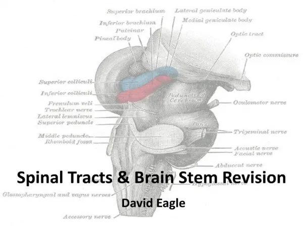
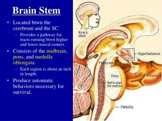
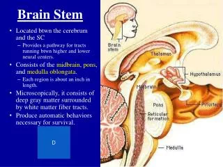
Brain stem. The brain stem. Consists of Midbrain Pons Medulla oblongata. 1. 2. 3. Medulla oblongata. Ventral surface Pyramid : contain pyramidal tract (corticospinal tract ) Decussation of pyramid : formed by crossing fibers of corticospinal tract

E N D
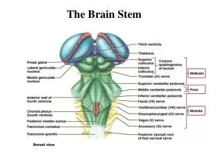
The brain stem Consists of • Midbrain • Pons • Medulla oblongata 1 2 3
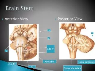
Medulla oblongata Ventral surface • Pyramid: contain pyramidal tract (corticospinal tract) • Decussation of pyramid: formed by crossing fibers of corticospinal tract • Olive: produced by underlying inferior olivary nucleus • Anterolateral sulcus: rootlets of hypoglossal nerve emerge from it • Retroolivary sulcus: rootlets of glossopharyngeal, vagus and accessory nerves emerge from it 1 IX XII 3 X XI 4 5 2
Medulla oblongata Dorsal surface Lower portion • Gracile tubercle:produced by underlying gracile nucleus • Cuneate tubercle: marks the site of cuneate nucleus • Inferior cerebellar peduncle • Obex Upper portion:forms the lower half of rhomboid fossa 3 5 2 4 1
Pons Ventral surface • Basilar part • Basilar sulcus • Bulbopontine sulcus: from medial to lateral, the abducent, facial and vestibulocochlear nerves appear • Middle cerebellar peduncle • Trigeminal nerve 1 V 5 2 VI 4 VII 3 VIII
Pons Dorsal surface • Superior cerebellar peduncle • Superior medullary velum • The upper half of rhomboid fossa 2 1
Midbrain Ventral surface • Crus cerebri • Interpeduncular fossa oculomotor nerves emerge from medial of crus cerebri • Posterior perforated substance • Oculomotor nerve 2 1 III 3 VI
Midbrain Dorsal surface • Superior colliculus constitute centers for visual reflexes • Inferior colliculus associated with auditory pathway • Brachium of superior colliculi • Brachium of inferior colliculi • Trochlear nerve 1 2 IV
Fourth ventricle Central canal →fourth ventricle →mesencephalic aqueduct→third ventricle Position • Situated ventral to cerebellum, and dorsal to pons and cranial half of medulla
Boundaries • Inferolateral: gracile and cuneate tubercles, inferior cerebellar peduncle • Superolateral: superior cerebellar peduncle • Lateral recess
Floor Upper pontine and lower medullary separated by Striae medullares Median sulcus divided it into righ andd left halves (2Medial eminence) Upper part: • Facial colliculus: overlies nucleus of abducent n. and fibers of facial nerve • Upper Vestibular area: overlies vestibular nuclei • Superior fovea
Lower part: • Hypoglossal triangle: verlying hypoglossal nucleus • Lower Vestibular area • Vagal triangle: overlies dorsal nucleus of vagus nerve • Inferior Fovea 2 2
Roof • Upper part: formed by superior cerebellar peduncle and superior medullary velum • Lower part: formed by inferior medullary velum and choroid plexus of fourth ventricle • Three apertures • Median aperture of fourth ventricle • Two lateral apertures of fourth ventricle