Download

1 / 59
690 likes | 878 Views
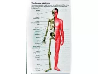
Imaging for Arthritis. NP/FP Outreach Curriculum in Rheumatology September 16, 2010 Dr. Sherry Rohekar. Outline. Introduction to imaging modalities Focus on plain radiography OA RA PsA AS Gout Pseudogout. X-rays. Taking a 2-dimensional image of a 3-dimensional structure

E N D
Imaging for Arthritis NP/FP Outreach Curriculum in Rheumatology September 16, 2010 Dr. Sherry Rohekar
Outline • Introduction to imaging modalities • Focus on plain radiography • OA • RA • PsA • AS • Gout • Pseudogout
X-rays • Taking a 2-dimensional image of a 3-dimensional structure • Superimposition of structures can thus obscure pathology • Quality is also affected by patient positioning, exposure techniques • Multiple views of the same area are useful • Good for: fractures, bone lesions, osteophytes, joint space narrowing, erosions, cysts
Computed Tomography (CT) • Also uses x-rays, but is superior than plain radiographs • Improved contrast • 3-D imaging • Attenuation of the x-ray beam travelling through tissues is measured from multiple angles • Substantially increased patient exposure to radiation when compared to plain films • Good for: fractures, subluxations, sclerosis, cystic bone lesions, evaluation of surgical hardware
Ultrasound (US) • Uses the interaction of sound waves with living tissue to produce an image • Doppler modes allow the determination of the velocity of moving tissues (i.e., blood flow) • User dependent, so requires an experienced technician who can make real-time measurements • Difficult to assess all planes • Good for: joint effusions, tenosynovitis, ganglia, erosions in RA, bursitis, tendonitis, and for guided injections/aspirations
Magnetic Resonance Imaging (MRI) • Based on the absorption and emission of energy in the radiofrequency range of the electromagnetic spectrum • No ionizing radiation exposure, superior soft tissue contrast resolution, excellent for the assessment of soft tissues, can image in multiple planes • Takes a long time to get access to scanner • Good for: tenosynovitis, joint effusions, synovial proliferation, cysts, erosions, cartilage loss, reactive bone changes
Nuclear Scintigraphy • In addition to showing anatomy, also provides information about underlying physiology • Most commonly used for MSK imaging: technitium-99m methylenediphosphate (Tc-MDP) • Can detect synovial hyperemia on the blood pool phase and periarticular uptake on the delayed phase in joints affected by inflammatory arthritis • VERY nonspecific, most rheumatologists consider the results to not be useful in clarifying the diagnosis • Good for: determining total number and symmetry of joints involved
Approach to an Image • Soft tissues: effusions, calcification, masses • Mineralization: diffuse demineralization, periarticular demineralization • Joint narrowing and subchondral bone: narrowing, subchondral sclerosis, intraarticular bodies, ankylosis • Erosions: central (articular surface), marginal (bare area), periarticular, mutilans • Proliferation: osteophytes, periostitis • Deformity: varus/valgus, flexion/extension, subluxation, dislocation, collapse • Distribution: monoarticular, pauciarticular, polyarticular, symmetric/asymmetric
Osteoarthritis • Joint space narrowing, osteophytes, subchondral sclerosis, cysts • Joint effusions are not uncommon • Early osteophytes look like sharpening of the joint edges • Distribution: weight bearing joints (hips, knees, back) • In the hands: DIPs, PIPs, CMC of thumb • Shoulder: glenohumeral OA usually secondary to rotator cuff disease
Rheumatoid Arthritis • RA characterized by synovial proliferation (pannus), bursitis and nodules • Can cause ill-defined soft tissue planes and prominances on plain films • Nodules appear as focal soft tissue masses especially at the olecranon bursa and areas of friction • Tenosynovitis can appear as diffuse soft tissue swelling, commonly seen at the wrist • Periarticular osteoporosis is an early finding , but can also see generalized osteoporosis
Rheumatoid Arthritis • Characteristic lesions are erosions in the marginal (bare) area • Pannus erodes the bone at the margin of the joint capsule where the redundant synovium exits, next to the articular cartilage • Osseous proliferation is not commonly seen with RA, but can be seen with secondary OA in joints with RA • Subchondral cysts may be large • Earliest changes are usually in the hands and feet • Ulnarstyloid soft tissue swelling, extensor carpiulnaristenosynovitis
Marginal erosion Erosions Soft tissue swelling
Rheumatoid Arthritis • Deformities • Subluxations at the MCPs and MTPs • Ulnar deviation of the digits • Swan-neck and Boutonniere deformities
Severe ulnar deviation Severe erosions of MCPs Complete destruction of the wrist Resorption of the carpals and the heads of the metacarpals Radial deviation of the wrist
Boutonniere deformity of the thumb Flexion with dislocation of the first MCP joint Hyperextension of the IP joint
Rheumatoid wrist: articular destruction, carpal fusion and carpal collapse. Severe destruction of the distal radius and ulna.
Rheumatoid foot Multiple osseous erosions and defects at the medial and lateral margins of the metatarsal heads Marginal erosions at the bases of the proximal phalanges (arrows)
Rheumatoid foot Medial and lateral erosions of the 5th metatarsal head Subluxation of the 5th MTP joint
Rheumatoid foot Subchondral cyst at the base of the distal phalanx Characteristic erosion along the medial margin of the proximal phalanx of the great toe
Soft tissue findings in rheumatoid knee Synovial effusion in the suprapatellar pouch and posterior recesses
Atlantoaxial subluxation in RA Always a concern in patient with longstanding RA and neck pain or cervical neurological symptoms
Order a view of the atlantoaxial articulation through an open mouth to fully assess. This shows lateral atlantoaxial subluxation of the odontoid process with respect to the lateral masses of the atlas.
Psoriatic Arthritis • Characterized by erosions and bony proliferations • RA does not typically have new bone formation • Asymmetric distribution • Typical “ray” distribution (involves several joints along a single digit) • Can involve the axial skeleton, as in ankylosingspondylitis (AS) • Soft tissue findings: fusiform soft tissue swelling around the joints; can progress so the whole digit is swollen (sausage digit or dactylitis) • “Fluffy” periostitis at the entheses • Marginal erosions also often show fluffy periostitis from new bone formation
Psoriatic Arthritis • Deformities • Pencil and cup – end of bone looks like it has been through a pencil sharpener, reciprocating bone becomes cupped • Telescoping of one bone into another • Complete destruction of bone (arthritis mutilans)
Psoriatic hands Erosive changes at the DIPs and PIPs Sparing of MCPs and wrists
Arthritis mutilans Pencil and cup deformity Pencilling
Psoriatic arthritis Asymmetric involvement Soft tissue swelling and periosteal reaction in 2nd and 3rd fingers
Psoriatic Arthritis • Spine • Asymmetric sacroiliitis • Chunky, asymmetrical syndesmophytes (bony bridges between vertebrae)
Chunky, non-marginal syndesmophytes typical of psoriatic arthritis
Asymmetric sacroiliitis with left sided erosions and sclerosis
Ankylosing Spondylitis • Changes begin at SI joints and lumbosacral junction, then typically move up the spine • SI joints: • Initially subchondral sclerosis • Then, small erosions cause “pseudowidening” of the SI joints • Erosions occur first at iliac side, which has thinner cartilage • Remember that the synovial part of the SI joint is the anterior, inferior portion • Reactive sclerosis with eventual fusion
Ankylosing Spondylitis • Spine • Early changes include squaring of the anterior vertebral body • Enthesitis (whiskering) and sclerosis (shiny corners) where Sharpey’s fibres mineralize • Progressive mineralization of Sharpey’s fibres to form osseous bridging syndesmophytes • Ossification of the interspinous ligaments • Most commonly involved peripheral joint is the hip
Erosions and sclerosis on iliac side Bilateral sacroiliitis with erosions, bony sclerosis and joint width abnormalities Bilateral sacroiliitis, definite erosions, severe juxta-articular bony sclerosis and blurring of the joint
Advanced AS Fused sacroiliac joints Ankylosis of the lower lumbar spine (bamboo spine)
Cervical spine in AS Shiny corners Squaring of the vertebral bodies Syndesmophytes
Gout • Erosions and masses, especially in the peripheral joints • Masses may be dense, due to crystals or associated calcification • Erosions are juxtaarticular from adjacent soft tissue tophi or intraosseous crystal deposition • Appear rounded with a well circumscribed sclerotic margin • Deformity occurs early • Olecranon and prepatellar bursitis may calcify