Download
1 / 15
150 likes | 307 Views
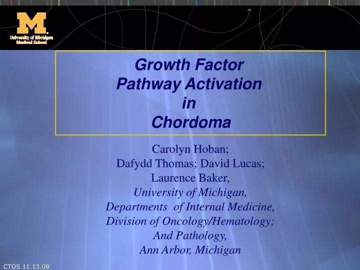
Growth Factor Pathway Activation in Chordoma. Carolyn Hoban; Dafydd Thomas; David Lucas; Laurence Baker, University of Michigan, Departments of Internal Medicine, Division of Oncology/Hematology; And Pathology, Ann Arbor, Michigan. Chordoma. Backgound:

E N D
Growth Factor Pathway Activation in Chordoma Carolyn Hoban; Dafydd Thomas; David Lucas; Laurence Baker, University of Michigan, Departments of Internal Medicine, Division of Oncology/Hematology; And Pathology, Ann Arbor, Michigan CTOS 11.13.08
Chordoma Backgound: • Tumor microenvironment of bone and nervous system • Usually slow growing tumors believed to arise from remnants of notochordal tissues • Challenging to treat with surgery, radiation and/or chemotherapy regimens due to location of the tumor. • Frequently recur and may have distant metastasis in late stage of disease. Factors regulating recurrence have not been determined. CTOS 11.13.08
Translational focus • To determine expression levels and activation of key growth factor pathways immunohistochemically using quantitative methods. • Retrospective analysis of routinely processed paraffin sections of chordoma clinical specimens. CTOS 11.13.08
Chordoma Clinical Specimens • 34 cases of Chordoma obtained from UM pathology archives with IRB approval • 3 tumor cores /pt /TMA • Diagnostic confirmation upon review by pathologists (Lucas) • M:F = 14:14; Age: avg. 52 (Range 11-81) • Anatomic location: • Clival (n=11); Sacral (n=9) • Primary and recurrent tumors CTOS 11.13.08
Low to intermediate-grade tumor, resembling notochord Clusters of large polymorphous cells in myxoid matrix Pleomorphic & hyperchromatic nuclei Mucin filled vacuoles ‘physaliferous’ Positive IHC for Cytokeratin19, S100, EMA Chordoma Histology CTOS 11.13.08
Brachyury*[Chordoma marker] RTK: HER family PDGFR and , IGF1R, c-Ret, c-kit, c-src, mTOR, AKT, and MAPK 2008 Tirabosco: Am J Surg Pathol, 2005 Henderson Genome Biology 2006 Vujovic J. Paht. IHC: Identification of activated pathways & drug targets CTOS 11.13.08
TMA 6971 TMA 6974 TMA 6983 Chordoma TMA: IHC scores Brachyury: high high frequency 100% high intensity 3+ • c-kit: neg/ low • HER family • EGFR mod • EGFRVIII neg • Her2: neg/ low TMA 8387 TMA 6971 1 2 3 Fasig et al. ckit low (33%); EGFR (67%) Weinberger et al. Her2 focal staining CTOS 11.13.08
PDGF-R PDGF-R TMA 6977 IHC scores: GF receptor profile • PDGF_R: high • c-ret: high (GDNF pathway) • IGF1R • Phospho-tyr IGF1R TMA 6985 pY1147-IGF1R IGF1R CTOS 11.13.08
Quantitative IHC • Automated Quantitative Analysis (AQUA) • Advantages: • Automated, high-throughput, • Objective (r/o intra/inter-observer concordance; lesion heterogeneity) • Fluorescence-based Ab used in IHC • Multiplex: • Antigen of interest • Tumor- and stromal-specific marker • Subcellular location (nuclei, cytoplasm,PM) • Automated scoring and data analysis • Activation index (phospho:total GF-receptor) CTOS 11.13.08
AQUA analysis Tumor Stroma Cytokeratin Cy3 Necrosis Tumor Mask Nuclear Component CTOS 11.13.08
IGF1R, c-ret, PDGFR pathways are activated AQUA score CTOS 11.13.08
From Clinical evidence to Preclinical models CTOS 11.13.08
In vitro models (chordoma cell lines) U-CH1 (Scheil et al 2001) [courtesy of Chordoma Foundation, Josh Sommer; Mike Kelly, Duke Univ) Morphological features: physaliferous, round nuclei, mucinous, (BrU,CK, S100 & EMA positive) Other cell lines in development In vivo models (chordoma/ microenvironment) UCH-1 cells with stable expression of luciferase Developing BLI- Xenograft lines (passage in SCID mice) Plan to evaluate drugs that inhibit GF pathways in vivo Preclinical models of chordoma CTOS 11.13.08
Conclusion Used data obtained from clinical specimens to build our experimental models: • Rank cell lines with similar features to clinical specimens (molecular markers, immunopositive GF pathway activation) • Test drug combinations to optimally inhibit key pathways, in vitro (cell kill) and in vivo (OS) • Support nomination of optimal combinations of drugs into clinical research • Translation Clinic-lab-Clinic CTOS 11.13.08
Thank you • The team: • University of Michigan Multidisciplinary Sarcoma group • Collaborators • Patients & friends (the village) Financial support from • Stefan L. Harris Fund Contact: carhoban@umich.edu CTOS 11.13.08