Download
1 / 24
240 likes | 426 Views
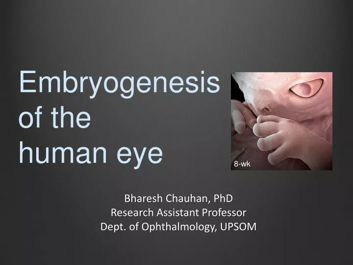
Embryogenesis of the human eye. 8-wk. Bharesh Chauhan, PhD Research Assistant Professor Dept. of Ophthalmology, UPSOM. Examples of inborn errors in human eye development. Aniridia. Anophthalmia. Coloboma. (Dr. K. Ramaesh , FRCS FRCOphth University of Edinburgh).

E N D
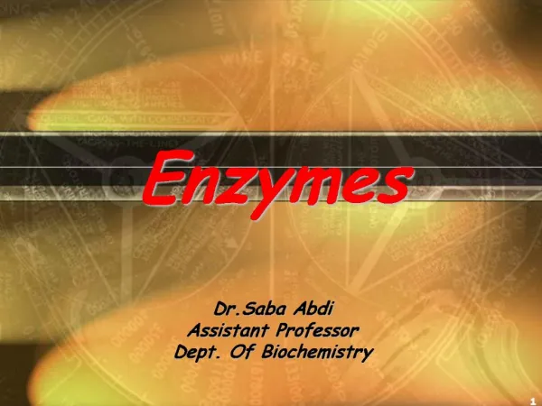
Embryogenesis of the human eye 8-wk Bharesh Chauhan, PhD Research Assistant Professor Dept. of Ophthalmology, UPSOM
Examples of inborn errors in human eye development Aniridia Anophthalmia Coloboma (Dr. K. Ramaesh, FRCS FRCOphth University of Edinburgh) (Dr. W.C. Caccamise, Sr, MD Pittsford, NY) (Dr. B.P. Brooks, MD PhD NEI, MD)
Stages in animal development Gametogenesis sperm (spermatogenesis) and ovum (oogenesis) form. Fertilization fusion of a sperm and ovum to generate a zygote. Cleavage to form morula (solid ball of blastomeres) and then blastula (ball of cells surrounding blastocoele). Gastrulation reorganization of blastocyst into a gastrula, consisting of endoderm, mesoderm and ectoderm. Organogenesis organ formation from the trilaminar gastrula through the processes of patterning, differentiation, morphogenesis and growth. Growth correct size and shape of organs are determined by growth in creating the fetus.
Organogenesis – the process Regional specification cell differentiation morphogenesis growth
Mammalian eye development Optic stalk The genetic and molecular basis of congenital eye defects. GrawJ. Nat Rev Genet. 2003 Nov;4(11):876-88. Kismet/CHD7 regulates axon morphology, memory and locomotion in a Drosophila model of CHARGE syndrome. MelicharekDJ, Ramirez LC, Singh S, Thompson R, Marenda DR. Hum Mol Genet. 2010 Nov 1;19(21):4253-64. Photoreceptor neurons Developing brain
Neurulation and head patterning • Prior to retinal development, the following occurs during the first 3 weeks of embryogenesis: • - neural induction of the competent ectoderm • - anterior-posterior subdivision of the resulting neural plate. Neural plate patterning: upstream and downstream of the isthmic organizer. Wurst W, Bally-Cuif L. Nat Rev Neurosci. 2001 Feb;2(2):99-108. Neural crest specification: migrating into genomics. Gammill LS, Bronner-Fraser M. Nat Rev Neurosci. 2003 Oct;4(10):795-805.
Placode stage • Specification of the eye field in the diencephalon occurs at this stage by signaling between the closely-apposed: • - epithelia of the surface ectoderm • - the lateral diverticuli of the budding diencephalon.
Lens pit stage • The first morphological sign of eye development in vertebrates is the bilateral evaginationof the diencephalon to form the optic vesicles. • Continued evagination leads to the formation of the optic pits with elongation of the optic stalk driving the back of the eye further. • Mesenchyme between the optic vesicle and the surface ectoderm is displaced as the two tissues come into close physical contact.
Optic cup stage • The continued evagination of the optic vesicle leads to the formation of the optic cup, which is bilayered. • The upper layers differentiate to form the neural retinas and the lower layers differentiate to form the retinal pigment epithelium (RPE).
Differentiation of RPCs into retinal cell types Intrinsically different retinal progenitor cells produce specific types of progeny. Cepko C. Nat Rev Neurosci. 2014 Sep;15(9):615-27. • human neural retina of the optic cup inner layer differentiates within first month • ganglion cells start first (7-8 wks) and centrally, while bipolar and horizontal cells start peripherally and migrate centrally, • primitive fovea is formed from the thinning of the GCL and INL at 24-26 wks .
Optic stalk (OS) – choroidal fissure closure • occurs at 6 weeks, • molecular mechanism has been partially deciphered (eg., FGF, RA).
Optic nerve development Four phases of development 1. 2. 3. • optic stalk extension • choroidal fissure closure (coloboma), • migration of the RGC axons into the optic stalk towards the LGN, • reduction of axons by apoptosis (megalopapilla) • Myelination of the axons occurs later in gestation, 32 wks in humans
Hyaloid vasculature development Three stage process In vivo analysis of hyaloid vasculature morphogenesis in zebrafish: A role for the lens in maturation and maintenance of the hyaloid. Hartsock A, Lee C, Arnold V, Gross JM. Dev Biol. 2014 Aug 13. pii: S0012-1606(14)00376-5. • deposition of hyaloid cells around • lens area and generation of a loop, • branched networks form, • vessel refinement occurs.
placode pit vesicle fiber a critical phase for co-ordinated morphogenesis between two epithelia, the optic vesicle and presumptive lens Four stages of morphogenesis in lens development
Mechanism of invagination Balanced Rac1 and RhoA activities regulate cell shape and drive invagination morphogenesis in epithelia. Chauhan BK, Lou M, Zheng Y, Lang RA. ProcNatlAcadSci U S A. 2011 Nov 8;108(45):18289-94. RhoA Rac1 •RhoA promotes apical constriction •Rac1 promotes cell lengthening
Mann I.(1950)The Development of the Human Eye. 2nd Ed. Grune & Stratton, Inc. New York. Inter-epithelial processes during lens placode invagination
Lens pit filopodia contain active myosin II Cdc42- and IRSp53-dependent contractile filopodia tether presumptive lens and retina to coordinate epithelial invagination. Chauhan BK, Disanza A, Choi SY, Faber SC, Lou M, Beggs HE, Scita G, Zheng Y, Lang RA. Development. 2009 Nov;136(21):3657-67.
Does the developing lens shape the optic cup? 1) • Shape of the cultured optic cup is different from the naturally developing optic cup next to the lens. • Uncoupled developing lens and optic cups do not conform to canonical shape forms. Self-organizing optic-cup morphogenesis in three-dimensional culture. Eiraku M, Takata N, Ishibashi H, Kawada M, Sakakura E, Okuda S, Sekiguchi K, Adachi T, Sasai Y. Nature. 2011 Apr 7;472(7341):51-6. 2) Cdc42- and IRSp53-dependent contractile filopodia tether presumptive lens and retina to coordinate epithelial invagination. Chauhan BK, Disanza A, Choi SY, Faber SC, Lou M, Beggs HE, Scita G, Zheng Y, Lang RA. Development. 2009 Nov;136(21):3657-67.
Maturation of the lens and sutures • Fiber cell differentiation begins during early vesicle stage to form primary fibers in the posterior lens, • lens continues enlarging by proliferation and elongation of the lens equator epithelial cells to form secondary fibers, • The fibers adopt a hexagonal shape, a ball-socket mid-arrangement and are bound to the lens capsule by a foot processes https://mutagenetix.utsouthwestern.edu/phenotypic/phenotypic_rec.cfm?pk=309 Understanding the role of growth factors in embryonic development: insights from the lens.LovicuFJ, McAvoy JW, de Iongh RU. PhilosTrans R SocLond B Biol Sci. 2011 Apr 27;366(1568):1204-18. doi: 10.1098/rstb.2010.0339. Review. Three-dimensional organization of primary lens fiber cells. Shestopalov VI, Bassnett S. Invest Ophthalmol Vis Sci. 2000 Mar;41(3):859-63.
Corneal development Neuroblastomabook edited by Hiroyuki Shimada, ISBN 978-953-51-1128-3, Published: May 29, 2013 Chapter 4 Neurotrophinand Neurotrophin Receptor Involvement in Human Neuroblastoma PierdomenicoRuggeri, Antonietta R. Farina, Lucia Cappabianca, Natalia Di Ianni, MarziaRagone, StefaniaMerolle, Alberto Gulino and Andrew R. Mackay
Angle development The next step: detailed assessment of an adult glaucoma patient. Philippin H, Shah P, Burton M. Community Eye Health. 2012;25(79-80):50-3.