Download
1 / 18
180 likes | 299 Views
The use of PCA-based Methods in the Design of Normal Spect rCBF atlases. Charlotte Bjuren Supervisors: Prof. Alex Houston Prof. Peter Hancock. Aim of PhD.

E N D
The use of PCA-based Methods in the Design of Normal Spect rCBF atlases. Charlotte Bjuren Supervisors: Prof. Alex Houston Prof. Peter Hancock
Aim of PhD. • The aim of my PhD is to investigate the potential of a multivariate method for computer aided detection of abnormalities in SPECT imaging • We know that most brains are physiologically different; using a standardised template as found brain imaging software only corrects for anatomical differences. • To address this problem a procedure has been proposed for producing normal atlases, that involves registering and normalising normal images and extracting the mean image plus an appropriate number of normal variants (Eigen images) using Principal Components Analysis Postgraduate Presentation
Medical Image Format MRI Images SPECT Images Postgraduate Presentation
Single Photon Emission Computed Tomography Postgraduate Presentation
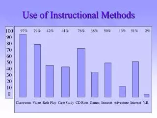
How Images are Processed. • Registration to template, (alignment of images) are done in Brass, or Multi-Modality. • Brass automatically fits images to an HMPAO templates, created from 35 individuals. • Displays Defect and Difference Images. • Multi-Modality can load up to 3 studies from different modalities. (Pet, CT, MRI & SPECT). • A good visualisation software is Hybrid Viewer. Postgraduate Presentation
EFFECT OF IMAGE REGISTRATION ON A SINGLE TRANSVERSE SLICE IMAGE SLICE TEMPLATE SLICE REGISTERED IMAGE SLICE MEAN IMAGE SLICE Postgraduate Presentation
Can’t tell individual from Mean Image! Postgraduate Presentation
MEAN IMAGE Postgraduate Presentation
EIGENIMAGE 1 Postgraduate Presentation
Extraction of Eigen Images. • Eigen vectors are formed by performing PCA on a set of Normal Images using the voxel as a variable. Since these eigenvectors are in the form of images, they are called eigen images. • They represent the ordered normal variants within the normal dataset and will include mainly physiological variation within the normal dataset and statistical noise. Postgraduate Presentation
Optimisation of the number of Eigen images. • The ration of variance contributed by an eigen image to the total variance in the normal dataset is given by its Eigen value.(eigenimages are ordered by their Eigen values) • It maybe assumed that signal-to-noise ratio in a Eigen image will decrease as the eigenvalue decreases. • An indication of how many Eigen images to include in the atlas is provided by a plot of Eigen values.(altough more sophisticated methods exist) Postgraduate Presentation
CUT-OFF AT P = 5 Postgraduate Presentation
First Year Project • The first year will involve developing a statistical method for determining the optimal number of Eigen images. • The method will then be applied to HMPAO brain SPECT-imaging. • We will compare various optimisation methods of extracting Eigen images such as an EigenvalueScree Plot vsJacknifing. Postgraduate Presentation
Reference List • Houston, A. S. (1998). Combining cross-validation and jackknifing to assess the validity of a normal brain atlas. In E. H. M. S. Berry (Ed.), (pp. 53-56). University of Leeds. • Houston, A. S., Kemp, P. M., Griffiths, P. T., & MacLeod, M. A. (1994a). An estimation of noise levels in HMPAO RCBF SPECT images using simulation and phantom data; comparison with results obtained from repeated normal controls. Physics in Medicine and Biology, 39, 873-884. • Houston, A. S., Kemp, P. M., & MacLeod, M. A. (1995). How can we define normality in a medical image? In Yves Bizais (Ed.), Information Processing in Medical Imaging: International Conference, Ile De Berder, France, June 1995 14th (Computational Imaging and Vision) (pp. 351-352). Kluwer Academic Publisher. • Houston, A. S., Fleming, J. S., Ward, T., & Hoffmann, S. M. A. (2009). Optimization of the parameters of a method for computer-aided detection of perfusion deficiencies in brain images. Nuclear Medicine Communications, 30. • Charlotte’s Homepage: charlottebjuren.com Postgraduate Presentation