Download
1 / 21
220 likes | 339 Views
External Beam Radiation Therapy Special Procedures. Kent A. Gifford, Ph.D., Acknowledgements to : Karl L. Prado, Ph.D., Charles Bloch, Ph.D., Peter Balter, Ph.D., Gurpreet Arora, B.S. Department of Radiation Physics. Total Body Irradiation. Indications Techniques Dosimetry Data Examples.

E N D
External Beam Radiation Therapy Special Procedures Kent A. Gifford, Ph.D., Acknowledgements to : Karl L. Prado, Ph.D., Charles Bloch, Ph.D., Peter Balter, Ph.D., Gurpreet Arora, B.S. Department of Radiation Physics
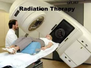
Total Body Irradiation • Indications • Techniques • Dosimetry Data • Examples
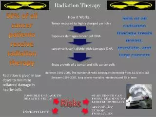
TBI: Clinical Indications • Preparation for marrow or stem-cell implantation • Suppress immune system to prevent rejection • Rid body of tumor cells not reached by chemotherapeutic agents (e.g. CNS)
TBI TechniquesAAPM Report 17, figure 1. source moves horizontally single source, long SSD half body, direct and oblique fields patient moves horizontally half body, adjacent direct fields
TBI TechniquesAAPM Report 17, figure 1. two vertical beams four sources single source, short SSD two horizontal beams head rotation
Total Skin Irradiation • High Dose-Rate Total Skin Electron (HDTSE-) Treatment: Technique Description • Indications • Methodology • Data / Calculation • Dose Verification (TLD)
MDACC TSEI technique(modified Stanford technique) • 2 angled beams (90°±23°) for uniform coverage and reduced total body dose • patient is 25 cm behind lucite plate • improved dose uniformity(6 fields w/plate 8 fields w/o plate, Holt JG, Perry DJ. Med. Phys.9:769-776(1982)) • increased dose to self shielded areas by diffusion • 6 positions, every 60°
st nd 1 day of cycle 2 day of cycle laser MDACC TSEI technique(modified Stanford technique) ANT POST RPO LPO LAO RAO
feet straight ahead or pointed slightly outwards for better balance elbows at or above shoulder level arms parallel to the scatter plate Stanford dual beam technique
QA - Patient TLD’s • TLD’s are taped to the patient’s skin and irradiated for one cycle (12 beams) • reference TLD’s are irradiated using a 10x10 cm2 field of 9 MeV electrons at2.0 cm depth (100 cm SSD) in anacrylic phantom
SRS: Available Systems- • Gamma Knife • Radionics Xknife/Xplan • Brainlab • Cyberknife • Others (many “homemade”)
Delivery techniques • 201 intersecting beams (Gamma Knife) • spherical dose distributions • Arcs with circular collimators (1-4 cm) • ellipsoid dose distributions • Multiple fields, shaped with miniature MLC • arbitrary dose distributions • IMRT
Elimination of motion & setup errors means smaller field size can be used Reduction in field size allows higher dose per fraction, and reduction in number of fractions (1 to 5) Large dose to small lesions with minimal dose to surrounding tissue. Single fraction “Out-of-town” patients Patients in poor health Other patients who might be burdened by protracted treatment. Stereotactic Radiosurgery Bloch, Ph.D.
Treatment • Typically 5 arcs, about 60 degree each. • 5-10 MU/degree 300 - 600 MU/arc. • 300 MU/min 1-2 minutes per arc. • Total treatment is 5-10 min of beam time. • Room entries between arcs to move couch and re-check patient alignment.
Gating Study-Left Breast By Gurpreet Arora, B.S.
Planning on the three CT sets CT at Inspiration CT at Expiration CT during Free Breathing Combined to form ITVs By Peter Balter, Ph.D. and Lei Dong, Ph.D.
The GE system for 4D scanning GE PET/CT (can be used on any GE CT scanner) Uses Varian RPMÔ to monitor respiration By Peter Balter, Ph.D.