Download
1 / 55
550 likes | 690 Views
Imaging signal transduction in single dendritic spines during synaptic plasticity. Ryohei Yasuda (HHMI, Duke). Spine. Spine: Biochemical compartment. Small ~0.1 fL. Narrow neck (~100nm Φ ) : Diffusional barrier Ca 2+ signaling in spines Synaptic plasticity Memory.

E N D
Imaging signal transduction in single dendritic spines during synaptic plasticity Ryohei Yasuda (HHMI, Duke)
Spine Spine: Biochemical compartment • Small ~0.1 fL. • Narrow neck (~100nmΦ): Diffusional barrier • Ca2+ signaling in spines Synaptic plasticity Memory
Signaling networks R. Iyenger
Imaging signaling in single spines • Measure FRET with 2-photon fluorescence lifetime imaging (2-photon FLIM) • Develop and use FRET sensors optimized for 2-photon FLIM • Image signal transduction, while inducing plasticity in single spine with 2-photon glutamate uncaging
Donor Mixture FRET FRET and fluorescence lifetime • FRET decreases fluorescence lifetime. • Use donor fluorescence only. • Independent of fluorophore concentrations. • Independent of wavelength-dependent light scattering • Multiple populations can be deconvolved. • Easy to combine with 2-photon microscopy Laser pulse Log (fluorescence) 2 4 6 8 10 12 Time (ns)
2-photon fluorescence lifetime imaging microscopy • High resolution and sensitivity in deep tissue. • Quantitative measurements of FRET
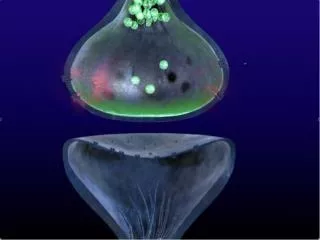
Stimulate single spines using 2-photon glutamate uncaging Matsuzaki, Ellis-Davies, Kasai Synapse-specific Ca2+ elevation
2p-uncaging to produce long lasting synaptic potentiation and spine growth 30-60 times, 0.5-1 Hz in Zero extracellular Mg2+ Matsuzaki 2001, 2004
Imaging activity ofCaMKIICa2+/Calmodulin-dependent kinase II
P Ca2+/Calmodulin-dependent kinase II: biochemical memory? Autoinhibitory domain CaM GFP Inactive Low Ca2+ d-YFP Kinase domain FRET Ca2+ Kinase domain Ac t i vation Ca2+/CaM Memory? Ca2+ P CaM Takao et al, 2004 Lisman 2002
CaM+, ATP+ CaM+, ATP– CaM–, ATP+ C a M– , A T P– C a M +, ATP+ (T286A) Fluorescence lifetime change in lysates Ca EG T A 2+ 0 . 5 0 . 4 0 . 3 Fluorescence lifetime change (ns) 0 . 2 0 . 1 0 - 5 0 5 1 0 1 5 Time (min) Lee et al., Nature 2009
CaMKII activation during structural plasticity of spines Lee et al., Nature 2009
Transient and spine-specific activation of CaMKII Lee et al., Nature 2009
Volume 400 300 200 Volume change (%) 100 0 -5 0 5 10 15 20 25 30 Time (min) Transient and spine-specific activation of CaMKII Uncaging 0.12 0.1 Uncaging 0.08 Stimulated Spine 0.06 Adjacent Spine CaMKII Activity Change Dendrite 0.04 0.02 0 ?0.02 -5 0 5 10 15 20 25 30 35 CaMKII 2 min 35 Lee et al., Nature 2009
Ca2+/Calmodulin-dependent kinase II: biochemical memory? Not for 1 hour CaM Inactive Low Ca2+ Ca2+ Kinase domain Ac t i vation P Ca2+/CaM Memory? Ca2+ P CaM What is the role of phosphorylation?
~0.1 s 5.8 s Wild-type 0.7 s T286A Fast fluorescence lifetime imaging Glutamate uncaging (4 ms) 0.3 1 μM 0.25 Δ[Ca2+] 0.2 0.15 Fluorescence lifetime (ns) 0.1 0.05 0 -0.05 -2 0 2 4 6 8 10 12 14 T ime (s) Lee et al., Unpublished
Ca2+/Calmodulin-dependent kinase II: biochemical memory? Yes, but only for 6 s CaM Inactive Low Ca2+ Ca2+ Kinase domain Ac t i vation P Ca2+/CaM Memory? Ca2+ ~ 6 s P CaM J. Lisman
0.1 s 10 s 1 min 10 min 1 hour Previous view of LTP Long-term plasticity CaMKII Ca2+
0.1 s 10 s 1 min 10 min 1 hour Now …. Long-term plasticity CaMKII Ca2+
Imaging the activity of Ras superfamily proteins
Small GTPase signaling • Several major subgroups: Ras, Rho, Rab, Rap, Arf, Ran etc… • Acts as signaling switch. • Regulate organization of actin cytoskeleton, membrane trafficking etc. • Important for morphogenesis of dendritic spines and plasticity • Mutations in the pathway are associated with mental retardation
Imaging binding between Ras and Ras binding domain (RBD) of Raf1 Ras CaMKII Yasuda et al., Nat.Neurosci. 2006 Harvey et al., Science 2008
0.1 s 10 s 1 min 10 min 1 hour AMPAR exocytosis Ras ERK Long-term plasticity CaMKII Ca2+
General approach to make sensors for Ras superfamily RhoA sensor Cdc42 sensor RhoA mGF P mGF P Cdc42 mRF P mRF P mRF P RTKN mRF P Pak3 8-89 a.a.. 65-118 a.a. WT S79A, F89A Cdc42 / RhoA: Important for regulation of actin cytoskeleton and dendritic spine morphology.
Making small GTPase sensors 1. Screen RXX Binding Domain in cuvette Kd ~ 1 – 5 uM for GTP form (RBD inhibits Rho inactivation) Kd > 50 uM for GDP form for low background 2. Test sensitivity & specificity in cell line 3. Test sensitivity & kinetics in neurons
Step1: Screen RBD and mutants FRET between GFP-CDC42 and PAK2-mCherry
Cdc42 activation is compartmentalized and sustained Stimulated spine Adjacent spine Hideji Murakoshi
RhoA activation spreads and sustained Uncaging Stimulated spine 10 Adjacent spine RhoA activation (%) 5 AP5 0 0 10 20 30 Time (min) Hideji Murakoshi
Spatial profile of Cdc42 Cdc42 Cdc42 10 8-56 s 1-5 min 5-10 min 10-20 min Spine 5 Before Binding fraction change (%) Dendrite 0 24 s 0 5 10 0 5 10 0 5 10 0 5 10 5 µm 2.15 ns 2.65 Distance (µm)
H-Ras H-Ras 20 6 min 2 min 2 min 15 10 Binding fraction change (%) 5 0 5 µm 15 0 10 20 0 10 20 5 15 5 Distance (µm) 2.2 ns 2.9 Spatial spreading of RhoA RhoA RhoA 15 8-56 s 1-5 min 5-10 min 10-20 min Spine 10 Dendrite Binding fraction change (%) Before 5 0 24 s 0 5 10 0 5 10 0 5 10 0 5 10 5 µm 2.0 ns 2.6 Distance (µm)
Effects of overexpression on length constant Rho A Rho A n = 20/18 n = 20/18 r = 0.16 r = 0.05 8 8 6 6 Length constant (µm) Length constant (µm) 4 4 2 2 0 0 0 2 4 6 8 0 50 100 [mEGFP-RhoA] ( µ M) [mCherry-RBD-mCherry] ( µ M)
Diffusion coupling at the spine neck PA-GFP tagged Ras * * * * *Constitutively active mutants Ras proteins: ~5 s CaMKII: ~60 s Lee, Harvey, Murakoshi
0.1 s 10 s 1 min 10 min 1 hour Cdc42 Long-term plasticity Ca2+ CaMKII RhoA
RhoA activation is CaMKII dependent Ctrl (stim) (CaMKII inhibitor) KN62 (NMDA receptor inhibitor) AP5 Uncaging Partial inhibition at early phase 10 RhoA activation (%) Late phase is CaMKII dependent 5 0 0 10 20 30 Time (min) Hideji Murakoshi
AP5 (NMDA receptor inhibitor) Uncaging 10 5 Cdc42 activation (%) 0 0 20 40 Time (min) Cdc42 activation is CaMKII dependent Control KN62 (CaMKII inhibitor) Hideji Murakoshi
0.1 s 10 s 1 min 10 min 1 hour Cdc42 Long-term plasticity Ca2+ CaMKII RhoA
Cdc42 is required for long-term structural maintenance Cdc42 Sustained growth Hideji Murakoshi
Cdc42 is required for long-term structural maintenance Cdc42 binding domain (24 hours) Volume change (%) Time (min) Cdc42 Sustained growth Hideji Murakoshi
RhoA is required for transient phase RhoA Transient growth Hideji Murakoshi
Stronger inhibition of RhoA inhibits both transient and sustained phases Volume change (%) Time (min) RhoA Transient/Sustained growth
RhoA/Cdc42 does not alter Ca2+-CaMKII Control C3: Rho inhibitor WASP: Cdc42 inhibitor CaMKII activation Ca2+ 0.15 0.15 0.1 0.1 CaMKII Lifetime change (ns) 0.05 0.05 0 0 -5 0 5 10 0 1 2 Time (min) Time (min) Cdc42 RhoA Structural plasticity 400 400 300 300 200 200 Volume change (%) Spine growth 100 100 0 0 -5 0 5 10 0 1 2 Time (min) Time (min)
Regulation of spine volume by Rho GTPases Uncaging 10 min CaMKII Volume Cdc42 RhoA Cdc42/RhoA CaMKII 2 min Spine growth Volume Cdc42 RhoA CaMKII Hideji Murakoshi
0.1 s 10 s 1 min 10 min 1 hour Cdc42 Long-term plasticity Ca2+ CaMKII Actin RhoA Transient plasticity Actin
0.1 s 10 s 1 min 10 min 1 hour AMPAR exocytosis Ras ERK Cdc42 PAK Long-term plasticity Ca2+ CaMKII Actin RhoA ROCK Transient plasticity Actin