Download

1 / 81
810 likes | 814 Views
8 Articulations. Section 1: Joint Design and Movement. Learning Outcomes 8.1 Describe the basic structure of a synovial joint, and describe common accessory structures and their functions.

E N D
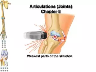
8 Articulations
Section 1: Joint Design and Movement Learning Outcomes 8.1 Describe the basic structure of a synovial joint, and describe common accessory structures and their functions. 8.2 Explain the relationship between structure and function for each type of synovial joint. 8.3 Describe flexion/extension, abduction/adduction, and circumduction movements of the skeleton. 8.4 Describe rotational and special movements of the skeleton.
Section 1: Joint Design and Movement Articulations (joints) Where two bones interconnect Bones are relatively inflexible so necessary to allow movement Reflect compromise between need for strength versus need for mobility Anatomical structure of each joint determines type and amount of movement possible Categories from range of motion and subgroups from anatomical structure
Section 1: Joint Design and Movement Three functional categories Synarthrosis (no movement) Amphiarthrosis (little movement) Diarthrosis (free movement) Synarthrotic and amphiarthrotic joints Relatively simple structure Direct connections between bones Diarthrotic joints Complex in structure Permit greatest range of motion
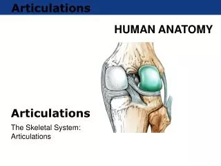
Module 8.1: Synovial joints Components of synovial joints Articular cartilages Resemble hyaline cartilages Matrix contains more water comparatively Have no perichondrium Slick and smooth, so reduce friction Are separated by thin film of synovial fluid
Module 8.1: Synovial joints Components of synovial joints (continued) Synovial fluid Similar in composition to ground substance in loose connective tissues Produced at the synovial membrane Circulates from areolar tissue to joint cavity Percolates through articular cartilages Total quantity is less than 3 mL
Module 8.1: Synovial joints Components of synovial joints (continued) Joint capsule Dense and fibrous May be reinforced with accessory structures (tendons and ligaments) Continuous with periosteum of each bone
Figure 8.1 1 The structure of synovial joints Medullary cavity Periosteum Components of Synovial Joints Articular cartilage Joint capsule Synovial fluid Synovial membrane Spongy bone of epiphysis Compact bone
Module 8.1: Synovial joints Functions of synovial fluid Lubrication With articular cartilage compression, synovial fluid is squeezed out and reduces friction between moving surfaces Nutrient distribution Provide nutrients and oxygen, as well as waste disposal for the chondrocytes of articular cartilages Compression and reexpansion of articular cartilages pump synovial fluid in and out of cartilage matrix Shock absorption Distributes compression forces across articular surfaces and outward to joint capsule
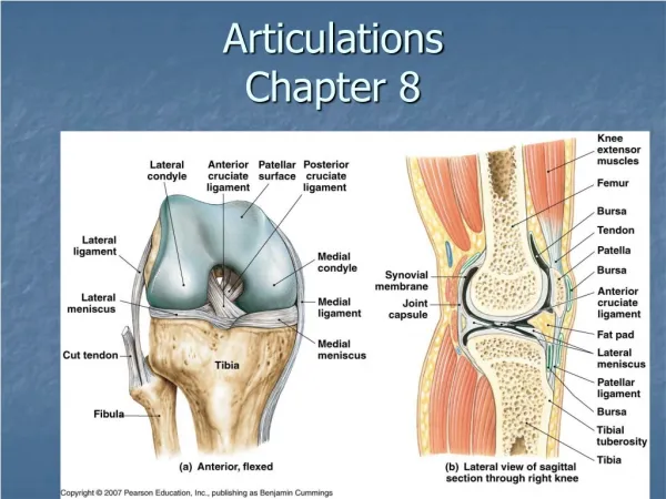
Module 8.1: Synovial joints Accessory structures Provide support and additional stability Not all are included in every joint Most are seen in the knee
Module 8.1: Synovial joints Accessory structures in knee Tendons of quadriceps Pass across joint Limit movement Provide mechanical support Bursa (a pouch) Small pocket filled with synovial fluid Often form in areas where tendon or ligament rubs against other tissues Reduce friction and act as shock absorbers
Module 8.1: Synovial joints Accessory structures in knee (continued) Fat pads Adipose tissue covered by synovial membrane Protect articular cartilages Act as packing material for joint Meniscus (a crescent) Pad of fibrous cartilage between bones of synovial joint May subdivide joint cavity and affect fluid flow or allow variations in shapes of articular surfaces
Module 8.1: Synovial joints Accessory structures in knee (continued) Accessory ligaments Support, strengthen, and reinforce joint Intrinsic ligaments Localized thickening of joint capsule Example: cruciate liagments of knee Extrinsic ligaments Separate from joint capsule May pass inside (intracapsular) or outside (extracapsular) the joint capsule Intracapsular example: cruciate ligaments Extracapsular example: patellar ligament
Figure 8.1 3 Accessory structures of complex synovial joints, as seen in a diagrammatic view of a sagittal section of the knee Tendon of the quadriceps muscles Patella Accessory Structures Synovial membrane Femur Bursa Joint capsule Fat pad Joint cavity Meniscus Articular cartilage Tibia Extracapsular ligament Intracapsular ligament
Module 8.1: Synovial joints Mobility vs. strength in joints Greater range of motion = weaker joint Examples: Synarthrosis (strongest type of joint, no movement) Diarthrosis (far weaker but broad range of motion) Dislocation (luxation) Movement beyond normal range of motion Articulating surfaces forced out of position Can damage joint structures No pain from inside joint but from nerves or surrounding structures
Module 8.1 Review a. Define a joint dislocation (luxation). b. Describe the components of a synovial joint, and identify the functions of each. c. Why would improper circulation of synovial fluid lead to the degeneration of articular cartilages in the affected joint?
Module 8.2: Types of motion and structural types of synovial joints Types of motion permitted at synovial joints Gliding Movement along two axes in one plane Angular motion Movement along two axes in one plane with additional change in angle
Module 8.2: Types of motion and structural types of synovial joints Types of motion permitted at synovial joints (continued) Circumduction Special complex angular movement Proximal end of bone remains fixed while distal end can move in a circle (“trace circumference”) Rotation Bone ends remain fixed and shaft rotates Animation: Synovial Joints: Movement
Figure 8.2 1 – 5 The general types of movement at synovial joints Starting position Gliding Angular motion Circumduction Rotation
Figure 8.2 6 The anatomical types of synovial joints, with joint models and examples Types of Synovial Joints Models of Joint Motion Examples Gliding joint • Acromioclavicular and claviculosternal joints Clavicle • Intercarpal and intertarsal joints Manubrium • Vertebrocostal joints • Sacro-iliac joints Hinge joint • Elbow joints • Knee joints Humerus • Ankle joints • Interphalangeal joints Ulna Pivot joint • Atlas/axis Atlas • Proximal radio-ulnar joints Axis Ellipsoid joint • Radiocarpal joints • Metacarpophalangeal joints 2–5 • Metatarsophalangeal joints Scaphoid bone Ulna Radius Saddle joint • First carpometacarpal joints Metacarpal bone of thumb Trapezium Ball-and-socket joint • Shoulder joints Scapula • Hip joints Humerus
Module 8.2 Review a. Identify the types of synovial joints based on the shapes of the articulating surfaces. b. What type of synovial joint permits the widest range of motion? c. Indicate the type of synovial joint for each of the following: shoulder, elbow, ankle, and thumb.
Module 8.3: Specific angular movements Flexion and extension Usually applied to movements of long bones of limbs but also axial skeleton Flexion Anterior/posterior movement that reduces angle between articulating elements Lateral flexion Vertebral column bending to the side Dorsiflexion Flexion at ankle joint and elevation of sole Plantar flexion (planta, sole) Extension at ankle joint and elevation of heel
Module 8.3: Specific angular movements Flexion and extension (continued) Extension Anterior/posterior movement that increases angle between articulating elements Hyperextension Extension past anatomical position Animation: Foot Dorsiflexion: Plantar Flexion Animation: Elbow Flexion/Extension Animation: Wrist Flexion/Extension
Figure 8.3 1 Flexion and extension Extension Flexion Hyperextension Lateral flexion Dorsiflexion (ankle flexion) Flexion Extension Plantar flexion (ankle extension) Hyperextension Flexion
Module 8.3: Specific angular movements Abduction and Adduction Always refers to movements of appendicular skeleton, not axial Movements are usually toward or away from body midline For fingers or toes, movements are spreading digits apart or bringing them together Abduction (ab, from) Movement away from body longitudinal axis in frontal plane Adduction (ad, to) Movement toward body longitudinal axis in frontal plane Animation: Humerus Abduction/Adduction
Figure 8.3 2 Abduction and adduction Adduction Abduction Abduction Adduction Abduction Abduction Adduction Adduction Abduction Adduction
Module 8.3: Specific angular movements Circumduction Moving arm or thigh as if to draw a big circle at distal end of limb Animation: Wrist Circumduction Animation: Humerus Circumduction Animation: Synovial Joints: Angular Movement
Module 8.3 Review a. When doing jumping jacks, which lower limb movements are necessary? b. Which movements are associated with hinge joints? c. Compare dorsiflexion to plantar flexion.
Module 8.4: Rotation and special movements Rotation When applied to the trunk, described as left and right rotation When applied to limbs Medial rotation (internal or inward rotation) Anterior surface of limb toward trunk long axis Lateral rotation (external or outward rotation) Anterior surface of limb away from trunk long axis Animation: Humerus Rotation
Figure 8.4 1 Rotational movements Left rotation Right rotation Lateral (external) rotation Medial (internal) rotation
Module 8.4: Rotation and special movements Rotation (continued) Other special terms for rotation of forearm Pronation Proximal end of radius rotates near ulna Distal end rolls across anterior ulnar surface Turns the wrist and hand from palm facing front to palm facing back Supination Opposing movement Palm is turned anteriorly Animation: Elbow Pronation/Supination
Figure 8.4 1 Rotational movements Supination Pronation
Module 8.4: Rotation and special movements Special movements Opposition Movement of thumb toward palm surface or other fingers Protraction Movement forward in anterior plane Retraction Reverse of protraction Inversion (in, into + vertere, to turn) Twisting foot motion to turn sole inward Eversion (e, out) Opposing movement to inversion
Module 8.4: Rotation and special movements Special movements (continued) Depression Movement inferiorly Elevation Movement superiorly Animation: Foot Inversion/Eversion Animation: Hand Opposition
Figure 8.4 2 Special movements Opposition Protraction Retraction Eversion Inversion Depression Elevation
Module 8.4 Review a. Snapping your fingers involves what movement with the thumb and third metacarpophalangeal joint? b. What movements are made possible by the rotation of the radius head? c. What hand movements occur when wriggling into tight-fitting gloves?
Section 2: Articulations Learning Outcomes 8.5 Describe the articulations between the vertebrae of the vertebral column. 8.6 Describe the structure and function of the shoulder and hip joints. 8.7 Describe the structure and function of the elbow and knee joints. 8.8CLINICAL MODULE Explain arthritis, and describe its effects on joint structure and function.
Section 2: Articulations Axial skeleton articulations Typically are strong but very little movement Appendicular skeleton articulations Typically have extensive range of motion Often weaker than axial articulations
Figure 8 Section 2 1 Joints of the Axial Skeleton Sutures of the skull Temporomandibular joint (temporal bone and mandible) Atlanto-occipital joint (occipital bone and atlas) and the atlanto-axial joint (C1–C2) Joints of the thoracic cage Intervertebral joints The lumbosacral joint, which attaches the last lumbar vertebra to the sacrum The sacrococcygeal and intercoccygeal joints, which structurally resemble simplified intervertebral joints
Figure 8 Section 2 2 Joints of the Appendicular Skeleton The sternoclavicular joint, the only articulation between the axial skeleton and the pectoral girdle and upper limb Shoulder joint The sacro-iliac joint, which firmly attaches the sacrum of the axial skeleton to the pelvic girdle of the appendicular skeleton Elbow joint Superior and inferior radio-ulnar joints Pubic symphysis Wrist joint Joints of the hand and fingers Hip joint Knee joint Ankle joint Joints of the foot and toes
Module 8.5: Vertebral articulations Vertebral articulations Between superior and inferior articular processes of adjacent vertebrae Gliding diarthrotic joints Permit flexion and rotation Adjacent vertebral bodies form symphyseal joints with intervertebral discs Numerous ligaments attach bodies and processes of vertebrae to stabilize column
Module 8.5: Vertebral articulations Intervertebral discs Composition Anulus fibrosis Tough outer layer of fibrous cartilage Collagen fibers attach to adjacent vertebrae Nucleus pulposus Soft, elastic, gelatinous core Provides resiliency and shock absorption Account for ¼ length of vertebral column Water loss from discs causes shortening of vertebral column with age and increases risk of disc injury
Figure 8.5 1 An intervertebral disc Anulus fibrosus Nucleus pulposus Superior view
Module 8.5: Vertebral articulations Primary vertebral ligaments Ligamentum flavum Connects adjacent vertebral laminae Posterior longitudinal ligament Connects posterior surfaces of adjacent vertebral bodies Interspinous ligament Connects spinous processes of adjacent vertebrae Supraspinous ligament Connects spinous processes from sacrum to C7 Ligamentum nuchae from C7 to base of skull Anterior longitudinal ligament Connects anterior surfaces of adjacent vertebral bodies
Figure 8.5 2 The ligaments attached to the bodies and processes of all vertebrae Primary Vertebral Ligaments Ligamentum flavum Intervertebral disc Anulus fibrosus Posterior longitudinal ligament Nucleus pulposus Spinal cord Interspinous ligament Spinal nerve Supraspinous ligament Posterior longitudinal ligament Anterior longitudinal ligament Sectional view Lateral view
Module 8.5: Vertebral articulations Disorders of vertebral column Slipped disc Posterior longitudinal ligaments weaken causing more pressure on discs Nucleus pulposus compresses, distorts anulus fibrosus Disc bulges into vertebral canal (doesn’t actually slip) Herniated disc Nucleus pulposus breaks through anulus fibrosus Spinal nerves are often affected
Figure 8.5 3 A slipped disc, as seen in a lateral view T12 Normal intervertabral disc L1 Slipped disc L2
Figure 8.5 4 A herniated disc, as seen in a superior view Compressed area of spinal nerve Spinal nerve Spinal cord Nucleus pulposus of herniated disc Anulus fibrosis
Module 8.5: Vertebral articulations Disorders of vertebral column (continued) Osteopenia (penia, lacking) Inadequate ossification leading to loss of bone mass Often occurs with age beginning between ages 30 and 40 More severe in women than men Osteoporosis (porosus, porous) Bone loss sufficient to affect normal function
Figure 8.5 5 The effects of osteoporosis on spongy bone Clinical scan of a compression fracture in a lumbar vertebra