Download
1 / 59
590 likes | 593 Views
Learn about the definition, causes, stages, and prevention of cancer, including the different types of tumors and warning signs to watch out for. Discover the importance of early diagnosis and ways to reduce your risk factors.

E N D
The Big “C”: CANCER By: Diana Blum RN MSN Metropolitan Community College
Definition • According to the American Cancer Society: • A large group of diseases characterized by uncontrolled growth and spread of abnormal cells
2nd leading cause of death in USA Metastasizing cancer Cell 1 out of 4 Americans will have CA at some time in their life
MEN Prostate Lung Colorectal Bladder Lymphoma Melanoma of Skin Oral Kidney Leukemia Stomach Women Breast Lung Colorectal Corpus Uteri Ovarian Lymphoma Melanoma of Skin Bladder Cervical Pancreas Common Sites
Men Lung Prostate Colorectal Women Lung Breast Colorectal Top 3 Cancers that cause Deaths
Normal Cell • Single small nucleus • Performs a specific function when it matures • Able to recognize other cells and identify tissue of origin • Reproduce in a controlled manner • Remain in their tissue of origin except blood cells
Neoplasm (aka TUMOR) • Cells that reproduce abnormally and in an uncontrolled manner
Benign Tumors • Harmless • Do not spread • Can create pressure or obstruct organs • 3 types • Fibroma: fibrous connective tissue • Lipoma: fat tissue • Leiomyoma: smooth muscle tissue
Malignant Tumors • Cancer cells characteristics • Change in appearance from normal cells/origin • Inability to properly perform function • Not recognized by other cells • Random disorganized uncontrolled growth pattern • Continue to divide when there’s no need • Can migrate to other organs
Malignant continued • Tend to press on normal tissue and organs as the grow • Invasive with all tissues • Regional invasion: movement into adjoining cells • Metastasis: to spread to distant sites • Most common sites are: • Treatment is more difficult with mets
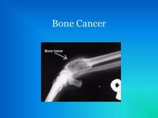
4 types of Malignancies • Carcinoma: skin, glands, lining of digestive urinary and reproductive tracts • Sarcoma: bone, muscle, other connective tissues • Melanomas: pigment cells in the skin • Leukemias and lymphomas: blood forming tissues: lymphoid tissue, plasma cells, and bone marrow
Malignant Transformation • 4 steps • Initiation: DNA exposed to carcinogen • Promotion: sufficient exposure to agent to encourage/enhance cell growth • Progression: accelerated growth, enhanced invasion, altered appearance and activity • Metastasis: tumor develops blood vessels • Penetrates capillaries and form fibrin network (undetectable by immune system) • Dissolve lining of blood vessels to invade surrounding tissue • Set up their own blood supply
Stage 1 • The malignant cells are confined to the tissue of origin. Not invasive with other tissues
Stage 2 • Limited spread of the cancer in the local area usually near lymph nodes
Stage 3 • The tumor is larger or has spread from the local site of origin into nearby tissues • regional lymph nodes are likely to be involved
Stage 4 • The cancer has metastasized to distant parts of the body
THE TNM Staging System • Specifies the status of the primary tumor, regional lymph nodes, and distant mets • T: tumor • N: regional nodes • M: distant mets
T=primary tumor T0: no signs of tumor after treatment Tis: malignancy in epithelial tissue but not in basement membrane T1: minimal size and extension T2, T3:progressive increase in size and extension T4:large size and extension N=regional nodes N0:no regional nodes involved N1:minimal node involvement N2:increased involvement of regional nodes N3:extensive involvement of regional nodes M=distant mets M0:no distant mets M1:distant mets present TNM Continued
practice • You are caring for a client who has cancer. The cancer is staged T4,N3, M1 how would you interpret the information??? • Primary tumor is large in size with extension that extensively involves the lymph nodes and distant mets are present.
Risk Factors • No single cause identified • Carcinogens exposure • (chemicals, radiation, viruses) • Cigarettes, asbestos, nitrates • Steroids, estrogens, tar, soot, asphalt, arsenic, corticosteroids, alkylating agents • Heredity and hormones also play factor • Familial cancers=appear at a high rate than expected. No single gene pinpointed • Heredity:predictable patterns of inheritance found on a single gene
7 warning signs of Cancer • C: change in bowel or bladder • A: a sore that does not heal • U: unusual bleeding or discharge • T: Thickening or lump in a breast or elsewhere • I: Indigestion or difficulty swallowing • O: Obvious change in a wart or mole • N: Nagging cough or hoarseness
Prevention and Detection • Health promotion • Avoid Carcinogens • ID high risk people
Health Promotion • Low fat, low cal, no added preservative, high fiber diet with at least 5 fruits and veggies daily • Avoid Alcohol • Avoid salt cured, smoked, or nitrate preserved foods • Balance activity and rest with stress management
Avoid carcinogens • Avoid over exposure from the sun • Do not smoke • Avoid second hand smoke • Wear a mask at work
ID High risk people • This helps researchers recognize factors that contribute to cancers • See doctor at least every 6 months
Diagnosis • H & P • Physical Exam • Diagnostic Tests • Biopsy: the removal of cells cut from a sample • Smear: blood cells under microscope to check for leukemia • CT Scan: used fto detect head and neck Ca, joints and soft tissue • MRI: detects CNS , spinal, neck, bones, joints, lung, kindey, etc. • PET(positron Emission Tomography): used to detect solid tumors in the brain and breast and to assess cancer treatment • Lab: Alpha fetoprotein , Ca50, Ca 125, PSA, etc. (see page 327)
Treatments • Surgery: • Done for: • diagnosis • Symptom relief • maintain function • Reconstruction • Possible cure
Surgery continued • Preop/postop care varies • The recommended treatment is based on the cancers: type, location, and mets
Radiotherapy • Uses ionizing radiation • Dose: 1 gray equals 100 rads • Used to treat malignant cells • Has delayed and immediate effects • Delayed: altered DNA which impairs the cells ability to reproduce • Immediate: cell death due to damage of cell membrane
Caregiver Safety with radiation • The less time spent near the source the less exposure • Unless direct care being given stay 6 ft away from the source • Effective shielding depends on type of rays (the denser the material the more protection)
External radiation • PROCEDURE • Source is outside the body • Special xray machine provides treatment • # of treatments depends on the doctor • Example: 5 times a week for 2-8 weeks • PATIENT PREP • Treatment simulation to determine exact dosage needed and schedule • The skin is marked with permanent, waterproof ink, by the radiologist for the exact site • Instruct client not to remove markings without permission
Internal Radiation (Brachytherapy) • PROCEDURE • Sources • Iodine, phosphorus, radium, iridium, radon, cesium • Instruct client that they pose a threat unitl the source is removed unless permanently implanted small beads used • 2 TYPES • Sealed • Unsealed
Sealed • Source is sealed in a container and inserted into the body (CESIUM) • Sources may be placed in threads, beads, needles, seeds, or molds • To protect visitors from exposure the client needs: • To be placed in isolation • Have a sign on the door indicating radiation • No pregnant women or kids under 18 allowed in room • Limit time with visitors • Have organized schedule for cares
Between scrotum and anus Intracavity such As bladder
Staff to wear film badges to monitor exposure • Recognize that Sealed sources can become dislodged • Portable lead shields provides minimal protection Immediately notify MD if source becomes dislodged. • Do not touch source with bare hands
Unsealed • Body fluids may be contaminated • Must wear gloves when working with patient • Contaminated fluids, dressings, etc may require additional precautions depending on the agency. • Disposable utensils are recommended • Equipment being removed from room must be checked for radiation level first
Radiation side effects • Normal cells may be harmed (hair follicles, bone marrow, lining of gi tractand urinary tract) • Anemia-deficiency of RBC • Low WBCs • Take 2-6 wks to recover • Bruising/Bleeding( low platelets) • Takes 2-6 wks to recover • Alopecia (hair loss) • Anorexia • Dry mouth • Harms reproductive cells
Nursing Implications • Teach pt to avoid exposure to sun, trauma, harsh chemicals, soaps • Teach pt to leave markings alone and to not use lotion during treatment • New hair may be different texture and color • Wig is okay to wear • Encourage dental care • Small frequent feedings • Monitor I/O • Increase fluid intake • Encourage C &DB
Chemotherapy • Use of chemical agents to treat (Antineoplastics) • Destroy rapidly dividing cells • Curative in some cases • Decreases symptoms in others
Antineoplastics (see Box 25-2) • Cell cycle phase specific- only works in a certain phase • Cell cycle phase non specific-works in all stages and phases • 5 types • Cytotoxic agents: Taxol, ifex, adriamycin, folex • Hormones and Hormone antagonists: femara, emcyt, evista • Biologic response modifiers: interferons, interleukins • Angiogenesis inhibitors: brand new and being studied
Chemo continued • Administered by doctor or certified nurse • Given inpt or outpt • Routes : po, iv, intracavity, or intrathecal • Intra cavity installed into cavity like bladder • Intrathecal is given in subarachnoid space • Perfusion:technique where drug is injected into artery supplying the tumor
Side Effects (see table 25-9) • Act on normal cells as well • Same as radiation • Bone marrow supression- most dangerous • N/V • alopecia • Client is also at risk for toxic effects • to heart (adriamycin)-causes heart failure • lung (Blenoxane)-pulmonary fibrosis and inflammation • nerve tissue (Velbane, Oncovin)- numbness, tingling, loss of deep tendon reflexes. • kidney, bladder
Biotherapy • Agents work by affecting biological processes including • hematopoietic growth factors (eyrthropoietin(production of RBC), numega, colony stimulating factors) • Biologic response modifiers (not first line treatment; still being studied), and • Monoclonal antibodies (specific for proteins on surface of cancer cell)
Transplants and hormone therapy • Bone marrow- used with leukemia/lymphoma • Stem cell- bone marrow depression • Both are done to restore blood manufacturing cells • Hormone therapy-used to supress natural hormone secretion, block hormone actions, or provide supplemental hormones
Nursing Assessment- diagnostic phase • Health History • Chief complaint, past medical hx, family history, system review (lumps, lesions, pain, fatigue, easy bruising, ha, hemoptysis, vision disturbance, loss of appetite, etc.(see pg 325) • Examination • Vs, ht, wt, inspect face, scalp, mouth for lesions • Ascultate lungs, and look at respiratory effort • Inspect breasts for symmetry, dimpling, lumps • Palpate abd, scrotum, etc