Download
1 / 54
0 likes | 10 Views
this is a PPt for parathormone and vit-Dits doses' clinical importance given in this slide

E N D
Parathormone and Vitamin-D By Dr. Vishnu Kumar
LEARNING OBJECTIVES • Introduction • Anatomy of the Parathyroid Glands • Regulation of Calcium Homeostasis • Chemistry of PTH • Biosynthesis of PTH • Mechanism of Action PTH • Metabolic role of PTH • Effects of PTH on the Body • Disorders Related to Parathormone • Diagnostic Tests for ParathormoneDisorders • Treatment Options • VITAMIN D • Sources of vitamin D • Metabolism and actions of vitamin D • Function of calcitriol • Clinical indications for vitamin D • Vitamin D Deficiency
Introduction • The parathyroid glands are intimately concerned with regulation of the concentration of Ca and PO4 ions in the blood plasma. • This is accomplished by secretion of a PTH by the chief cells, the net effect of which is: 1. To increase the concentration of Ca ↑ and Decrease the PO4 ↓ 2. In addition to its effects on plasma ionised Ca via its action on bone, parathormone controls renal excretion of Ca and PO4.
Anatomy of the Parathyroid Glands • The parathyroid glands are small endocrine glands located in the neck, situated close to the thyroid gland. • There are four parathyroid glands, and they are typically found in pairs on the back surface of the thyroid gland. • The two upper parathyroid glands are referred to as the superior parathyroids, and the two lower ones are called the inferior parathyroids. • Each parathyroid gland is about the size of a grain of rice, measuring approximately 3-8 mm in diameter. • The parathyroid glands are highly vascularized, allowing them to efficiently release parathormone (PTH) into the bloodstream when necessary. • PTH, as mentioned earlier, regulates calcium and phosphate levels by influencing the bones, kidneys, and intestines.
Regulation of Calcium Homeostasis • The regulation of calcium homeostasis by PTH involves several key mechanisms: • Stimulus for PTH Release: Low blood calcium levels act as the primary stimulus for the secretion of PTH. • The parathyroid glands are highly sensitive to changes in blood calcium concentrations. • PTH Secretion: The parathyroid glands, synthesize and store PTH. • When stimulated, they release PTH into the bloodstream. • Action on Bones: PTH acts on bones, which serve as the body's calcium reservoir. • It stimulates the process of bone resorption, where calcium and phosphate are released from the bone matrix into the bloodstream. • Effects on Kidneys: PTH influences the kidneys to increase calcium reabsorption from the urine back into the bloodstream. • This reduces the loss of calcium through urine and helps to conserve calcium in the body.
Chemistry of PTH • Parathormone is a linear polypeptide consisting of 84 amino acids. • N-terminal amino acid is alanine and C-terminal is glutamine. • Bovine PTH has molecular wt. of 9500. • Parathormone from different species differ only slightly in structure
Cont. • Core of Activity: the synthetic PTH indicate that the amino acid sequence 1 to 29 or possibly 1 to 34 from N-terminal end is essential for the physiologic actions of this hormone on both skeletal and renal tissues. • Methionine is important amino acid and necessary for calcium mobilising effect. • The N-terminal end up to 34 amino acids possesssthe “receptor-binding” ability.
PTH thus formed is packaged and stored in secretory vesicles. Increased c-AMP concentration and a low Ca++ level stimulates its release from secretory vesicles. On the other hand, a high concentration of Ca++ stimulates the degradation of the stored PTH in secretory vesicles instead of its release
Mechanism of Action • The mechanism of action of parathyroid hormone (PTH) involves several steps that ultimately lead to an increase in serum calcium (Ca++) levels. • PTH acts on bones, kidneys, and intestines to regulate calcium homeostasis in the body. (a) Increasing Cyclic AMP Level: PTH binds to specific receptors on the plasma membrane of target cells, such as bone cells and renal tubule cells. • This binding activates the enzyme adenylcyclase within these cells. • Adenylcyclase catalyzes the conversion of ATP to cyclic AMP (cAMP), which acts as a "second messenger" inside the cells.
Cont. (b) Role of Ca++: The increased cAMP levels in the cells also lead to an increase in intracellular calcium (Ca++) concentration. • This occurs because cAMP can modulate the activity of certain ion channels in the cell membrane, leading to an influx of calcium ions. • The increased intracellular calcium, in turn, affects various intracellular processes and signaling pathways.
Cont. (c) pH change in Tissues: PTH has been shown to increase the amounts of both lactic acid (LA) and citric acid in the tissues. • These acids may contribute to bone resorption, which is the process by which bone tissue is broken down and calcium is released into the bloodstream. • They are thought to play a role in the dissolution of hydroxyapatite, a major component of bone mineral.
METABOLIC ROLE OF PTH • The actions of PTH are reflected in the consequences of: • Its administration and Removal of the parathyroid glands. A. The most conspicuous metabolic consequences of administration of PTH are: • Increase in serum Ca++ concentration ↑ . • Decrease in serum inorganic PO4 ↓ concentration. • Increased urinary Ca++ ↑ following an initial decrease. • Increased urinary PO4 ↑ . • Removes Ca from bones, particularly if dietary intake of Ca is inadequate. • Increase in ‘citrate’ ↑ content of blood plasma, kidney and bones.
Cont. • Activates vit D in renal tissue by increasing the rate of conversion of 25-OH-cholecalciferol to 1,25-di-OH-cholecalciferol, by stimulating α-1- hydroxylase enzyme • Effect on Mg metabolism: PTH has been reported to exert an influence on Mg metabolism. • Primary hyperparathyroidism has been found to be associated with excessive urinary excretion of Mg and –ve Mg balance.
PTH Actions on Different Organs • Effects on Bones: • PTH stimulates bone resorption. • PTH stimulates osteoclasts, which are specialized cells responsible for bone resorption. • Osteoclasts break down bone tissue, releasing calcium and phosphate into the bloodstream.
PTH Actions on Different Organs cont.. • Effects on Kidneys: • PTH enhances calcium reabsorption in the distal tubules of the nephrons. • PTH signaling increases the expression of a calcium transporter called TRPV5, leading to increased calcium reabsorption from the urine back into the bloodstream. • PTH also inhibits phosphate reabsorption in the proximal tubules of the nephrons, leading to increased phosphate excretion in urine.
PTH Actions on Different Organs cont.. • Effects on Intestines: • PTH indirectly influences calcium absorption in the intestines through its activation of vitamin D. • PTH stimulates the conversion of inactive vitamin D (calcidiol) into its active form (calcitriol) in the kidneys. • Calcitriol, in turn, promotes calcium absorption from the intestines.
Disorders Related to Parathormone • Hyperparathyroidism: Causes, symptoms, and complications • Hypoparathyroidism: Causes, symptoms, and complications
Hyperparathyroidism • Hyperparathyroidism: Causes, symptoms, and complications. • Causes: • Hyperparathyroidism occurs when there is excessive secretion of Parathormone (PTH). • The most common cause of primary hyperparathyroidism is the development of a benign tumor (adenoma) in one of the parathyroid glands. • Less commonly, hyperplasia of multiple parathyroid glands or, rarely, parathyroid cancer can lead to hyperparathyroidism.
Hyperparathyroidism • Symptoms: • The condition is often asymptomatic, especially in mild cases. • However, some individuals may experience symptoms such as fatigue, weakness, bone pain, kidney stones, frequent urination, abdominal pain, and gastrointestinal disturbances. • Elevated blood calcium levels are a hallmark of hyperparathyroidism and can lead to various systemic effects.
Hyperparathyroidism • Complications • Long-term untreated hyperparathyroidism can result in serious complications eg. Osteoporosis (weakening of bones), increased risk of fractures, kidney damage (nephrolithiasis and nephrocalcinosis), and peptic ulcers. • It can also lead to hypercalcemic crisis, a severe and potentially life-threatening condition. • Very high calcium levels, causing altered mental status and scardiacabnormalities.
Hypoparathyroidism Causes: • Hypoparathyroidismoccurs when there is insufficient secretion of PTH from the parathyroid glands. • The most common cause of hypoparathyroidism is accidental damage or removal of the parathyroid glands during thyroid surgery. • Other causes include autoimmune diseases, genetic disorders, radiation therapy to the neck, or certain medications.
Hypoparathyroidism • Symptoms: • Hypoparathyroidismleads to low blood calcium levels and high blood phosphate levels. • Symptoms may include muscle cramps, twitching, spasms (tetany), numbness or tingling in the extremities. • Seizures, cognitive and mood changes, and dry, coarse skin
Hypoparathyroidism • Complications • If left untreated, hypoparathyroidism can lead to complications such as chronic kidney disease due to calcium deposits in the kidneys (nephrocalcinosis). • Cataracts, and abnormal dental enamel formation. • In severe cases, hypoparathyroidism can result in life-threatening low calcium levels and acute hypocalcemic crisis.
Diagnostic Tests for Parathormone Disorders • Blood tests to measure PTH and calcium levels • Imaging techniques for parathyroid gland evaluation
Blood tests to measure PTH and calcium levels Blood Tests: • PTH (Parathormone) Level: Measurement of PTH levels in the blood is a key diagnostic test for parathyroid disorders • common laboratory techniques used to measure parathyroid hormone (PTH) levels- • Immunoassays: • Radioimmunoassay (RIA): It involves using radioactive labels to measure the binding of antibodies to PTH molecules in a patient's serum. • Enzyme-Linked Immunosorbent Assay (ELISA): ELISA is a widely used technique that uses enzymes to generate a color change or signal when a specific antibody-antigen reaction occurs.
Cont. • Chemiluminescent Immunoassay (CLIA): • Principle of CLIA:Chemiluminescence involves the emission of light as a result of a chemical reaction. • In the case of CLIA, the reaction occurs between a substance of interest (in this case, PTH) and specific antibodies labeled with a chemical that emits light when triggered. • Fluorescence Immunoassay: This technique involves using fluorescent labels instead of radioactive or enzymatic labels. It relies on the detection of fluorescence signals emitted when labeled antibodies bind to PTH molecules.
Cont. • Electrochemiluminescence Immunoassay (ECLIA): ECLIA is a variation of CLIA that uses an electric current to stimulate the chemiluminescent reaction. • It is often used in automated immunoassay systems. • Liquid Chromatography-Mass Spectrometry (LC-MS/MS): This method is considered to be a more advanced and precise technique for measuring PTH. • It involves separating PTH from other components in the sample using liquid chromatography and then quantifying it using mass spectrometry. • LC-MS/MS can provide accurate measurements of various forms of PTH.
Cont. • Immunoradiometric Assay (IRMA): IRMA is another immunoassay method that uses radioactive labels. It measures the binding of antibodies to antigens • Intact PTH (pg/mL): Approximately 10 to 65 pg/mL
Calcium Level: Blood calcium levels are also measured to assess calcium homeostasis. In hyperparathyroidism, blood calcium levels are typically elevated, and in hypoparathyroidism, they are decreased. • Ionized Calcium Level: Ionized calcium is preferred over total calcium levels in specific cases, as it represents the physiologically active, free form of calcium in the bloodstream. • Phosphate Level: • Measurement of phosphate levels in the blood is important to assess its relationship with calcium levels. • Hyperparathyroidism often leads to decreased phosphate levels, while hypoparathyroidism is associated with increased phosphate levels. • Vitamin D Levels: • Vitamin D levels may be checked, especially in cases of hypoparathyroidism, as PTH plays a role in activating vitamin D to regulate calcium absorption from the intestines.
Imaging techniques for parathyroid gland evaluation • Ultrasound (US): Ultrasound is a non-invasive imaging technique that can help visualize the parathyroid glands and identify any abnormal growths. • SestamibiScintigraphy: This nuclear medicine scan involves the injection of a radioactive tracer called technetium-99m sestamibi, which is taken up by parathyroid tissue. • Computed Tomography (CT) Scan: CT scans can be used to provide detailed images of the neck and chest, and parathyroid tissue.
Magnetic Resonance Imaging (MRI): MRI may be used to assess the parathyroid glands and surrounding structures, providing additional information for surgical planning in cases of suspected parathyroid disorders. positron emission tomography (PET) were being investigated to improve the preoperative localization of abnormal parathyroid tissue.
PTHrP (Parathormone-related Peptide) • Also called as Humoralhypercalcaemic factor of malignancy (HHFM) • It is a peptide containing 141 amino acids. • Amino acid sequence on first 13 same (8 of 13 are homologous) from N-terminus • It is produced by a number of tumours specially squamous cells carcinomas of lungs, oesophagus, cervix and head and neck. • Also produced by renal carcinoma, carcinoma of pancreas, breast carcinoma, etc. • PTHrP can bind to parathormone receptor and can mimic the action of PTH. • Target tissues are bones and kidneys and produces hypercalcaemia, hypophosphataemia like PTH and also increases urinary cyclic AMP. • PTHrP is produced by a gene on chromosome 12 which is distinct from PTH gene which is located on chromosome 11.
CLINICAL IMPORTANCE OF PTHrP • Serum level of PTHrP are low or absent in normal healthy persons and in patients with primary hyperparathyroidism but it is high in majority of patients in malignancy and is responsible for humoralhypercalcaemia of malignancy. • Determination of serum PTHrP is becoming an important diagnostic tool in evaluation of hypercalcaema.
1. Medical Management of Hyperparathyroidism • Vitamin D and Calcium Supplements: In some cases, individuals with hyperparathyroidism may require vitamin D and calcium supplements, especially if they have vitamin D deficiency or low calcium levels due to the disease. 2. Surgical Interventions for Hyperparathyroidism: • Parathyroidectomy:Parathyroidectomy is the surgical removal of the affected parathyroid gland(s). • Minimally Invasive Techniques: Such as focused or targeted parathyroidectomy using preoperative localization studies (ultrasound or sestamibi scan), have become more common. • These techniques aim to remove the abnormal gland(s) while preserving the normal ones.
1. Medical Management of Hypoparathyroidism • Calcium and Vitamin D Supplements • Active Vitamin D (Calcitriol) or Analogs • Magnesium Monitoring: Magnesium levels should be monitored, as magnesium deficiency can impact PTH secretion and action. • Phosphate Management: Hypoparathyroidism can lead to elevated serum phosphate levels, which can further lower calcium levels. • Lifestyle and Diet: Patients are often advised to follow a diet that is adequate in calcium and low in phosphate. Dairy products, leafy greens, and fortified foods are good sources of dietary calcium. • Emergency Treatment: In severe cases of hypoparathyroidism, where the patient's calcium levels drop significantly (hypocalcemic crisis), intravenous calcium may be required to rapidly raise calcium levels to a safe range.
2. Surgical Interventions for Hypoparathyroidism 2. Surgical Interventions for Hypoparathyroidism: • Parathyroid Autotransplantation: Parathyroid autotransplantation involves re-implanting parathyroid tissue into another area, such as the forearm, to restore parathyroid function. • Parathyroid Gland Regeneration: Research is ongoing to explore the potential of stimulating the regeneration of damaged or non-functional parathyroid glands through various approaches, including stem cell therapies.
VITAMIN D • The D vitamins are a group of sterols that have a hormone-like function. • The active molecule, 1,25-dihydroxycholecalciferol ([1,25-diOH-D3], or calcitriol), binds to intracellular receptor proteins. • The 1,25-diOH-D3–receptor complex interacts with response elements in the nuclear DNA of target cells in a manner similar to that of vitamin A and either selectively stimulates or represses gene transcription. • The most prominent actions of calcitriol are to regulate the serum levels of calcium and phosphorus
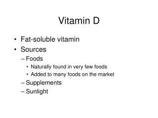
Sources of vitamin D • Ergocalciferol(vitamin D2), found in plants, and cholecalciferol (vitamin D3), found in animal tissues, are sources of preformed vitamin D activity. • Vitamin D2 and vitamin D3 differ chemically only in the presence of an additional double-bond and methyl group in the plant sterol
Metabolism • 1,25-Dihydroxycholecalciferol formation: Vitamins D2 and D3 are not biologically active but are converted in vivo to calcitriol. The active form of vitamin D - • The first hydroxylation occurs at the 25 position and is catalyzed by a specific 25-hydroxylase in the liver. • The product of the reaction, 25- hydroxycholecalciferol ([25-OH-D3], calcidiol), is the predominant form of vitamin D in the serum and the major storage form. • 25-OH-D3 is further hydroxylated at the 1 position by 25-hydroxycholecalciferol 1- hydroxylase found primarily in the kidney, resulting in the formation of 1,25-diOH-D3 (calcitriol).
Cont. 2. Hydroxylation regulation: • Calcitriol is the most potent vitamin D metabolite. • Its formation is tightly regulated by the level of serum phosphate and calcium ions . • 25- Hydroxycholecalciferol 1-hydroxylase activity is increased directly by low serum PO4 3− or indirectly by low serum Ca2+, which triggers the secretion of parathyroid hormone (PTH) from the chief cells of the parathyroid gland. PTH upregulates the 1-hydroxylase. • Thus, hypocalcemia caused by insufficient dietary Ca2+ results in elevated levels of serum 1,25-diOH-D3. • [Note: 1,25-diOH-D3 inhibits expression of PTH, forming a negative feedback loop. It also inhibits activity of the 1-hydroxylase].
Function of calcitriol The overall function of calcitriol is to maintain adequate serum levels of Ca2+. It performs this function by • Increasing uptake of Ca2+ by the intestine • Minimizing loss of Ca2+ by the kidney by increasing reabsorption 3) Stimulating resorption (demineralization) of bone when blood Ca2+ is low . 1. Effect on the intestine: Calcitriol stimulates intestinal absorption of Ca2+ by first entering the intestinal cell and binding to a cytosolic receptor. • The 1,25-diOH-D3–receptor complex then moves to the nucleus where it selectively interacts with response elements on the DNA.
As a result, Ca2+ uptake is enhanced by increased expression of the calcium-binding protein calbindin. Thus, the mechanism of action of 1,25-diOH-D3 is typical of steroid hormones . 2. Effect on bone: When blood Ca2+ is low, 1,25-diOH-D3 stimulates bone resorption by a process that is enhanced by PTH. The result is an increase in serum Ca2+. [Note: PTH and calcitriol also work together to prevent renal loss of Ca2+.]
Clinical indications for vitamin D • Nutritional rickets: Vitamin D deficiency causes a demineralization of bone, resulting in rickets in children and osteomalacia in adults . • In osteomalacia, demineralization of preexisting bones increases their susceptibility to fracture. • Insufficient exposure to daylight and/or deficiencies in vitamin D consumption occur predominantly in infants and the elderly. 2. Renal osteodystrophy: Chronic kidney disease causes decreased ability to form active vitamin D as well as increased retention of PO4 3−, resulting in hyperphosphatemia and hypocalcemia. • The low blood Ca2+ causes a rise in PTH and associated bone demineralization with release of Ca2+ and PO4 3−.
Vitamin D Deficiency • Supplementation: Vitamin D deficiency is commonly treated with oral vitamin D supplements. • The type and dosage of the supplement depend on the severity of the deficiency. • The two main forms of vitamin D used for supplementation are vitamin D2 (ergocalciferol) and vitamin D3 (cholecalciferol). • Dosage: The dosage can vary depending on factors such as the patient's age, underlying health conditions, and the extent of the deficiency.
Vitamin D Excess (Vitamin D Toxicity) • Discontinuation: If vitamin D levels become excessively high, vitamin D supplements should be discontinued. • Fluids and Medications:supportive measures such as intravenous fluids and certain medications may be used to help lower calcium levels and alleviate symptoms. • Underlying Conditions: Some medical conditions can interfere with the body's ability to process or utilize vitamin D. • Treating these underlying conditions can help improve vitamin D levels.