Download
1 / 1
10 likes | 138 Views
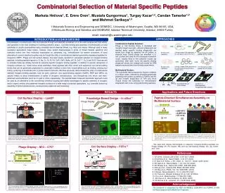
Au 0. 20mm. Glass. Glass. PDMS stamp. Glass. Glass. Fusion Protein. Keq (10 6 M -1 ). G (kcal/mol). KD (mM). l-AuBP1. 3.24±1.31. -8.9±0.2. 0.307. c-AuBP1. 2.51±0.50. -8.7±0.1. 0.398. l-AuBP2. 2.34±0.34. -8.7±0.1. 0.427. c-AuBP2. 13.50±3.00. -9.7±0.2. 0.074. Controls.

E N D
Au0 20mm Glass Glass PDMS stamp Glass Glass Fusion Protein Keq (106 M-1) G (kcal/mol) KD (mM) l-AuBP1 3.24±1.31 -8.9±0.2 0.307 c-AuBP1 2.51±0.50 -8.7±0.1 0.398 l-AuBP2 2.34±0.34 -8.7±0.1 0.427 c-AuBP2 13.50±3.00 -9.7±0.2 0.074 Controls l-AuBP2 l-AuBP1 c-HABP 100nm c-AuBP1 c-AuBP2 Citrate Peptide-Mediated Formation and Assembly of Gold Nanostructures Marketa Hnilova1, Alisa Carlson1,2, Chris, So1, Hanson Fong1, Candan Tamerler1,3 and Mehmet Sarikaya1,3 1 Materials Science and Engineering and GEMSEC, University of Washington, Seattle, WA 98195, USA, 2 Microbiology, University of Washington, Seattle, WA 98195, USA 3 Molecular Biology and Genetics and MOBGAM, Istanbul Technical University, Istanbul, 34469 Turkey email: markeh@u.washington.edu APPROACH INTRODUCTION and BACKGROUND First, we demonstrate the utility of the bifunctional QBP-AuBP peptide as a molecular linker to direct the immobilization of gold nanoparticles onto the silica glass surface using self-assembly and soft lithography techniques (micro-contact printing). Then, we used the silica glass decorated with gold nanoparticles for subsequent peptide-mediated gold reduction of chloroauric acid. As evidenced by dark field optical microscopy (DF) and atomic force microscopy (AFM), we selectively immobilized the gold nanoparticles via bifunctional QBP-AuBP peptide onto the silica glass. The hybrid composite nanostructures with controlled optical properties, especially gold-silica nanoshells, have a wide spectrum of biological applications in nanotechnology and medicine, such as biosensing, targeting, and bioimaging. Conventionally, the gold nanoshells are produced in a multistep process involving: i) the silica particle functionalization with organosilane molecules followed by ii) chemical coupling of gold nanoparticles to the silane molecules on silica nanoparticle surface and iii) by subsequent reduction of chloroauric acid by sodium borohydrate. Our research focuses on the benefits of genetically engineered polypeptides for inorganics (GEPIs) that can control both assembly and formation of various nanostructures. The gold- and quartz-binding peptide (AuBP and QBP) motives were selected from combinatorial peptide libraries in directed or in silico evolution. Their material specificity to given material versus other noble metals and silica was demonstrated in various studies including fluorescent microscopy (FM) and surface plasmon resonance (SPR). Here we combined these to motives and created a novel bifunctional peptide: QBP1 BF Pt Au 10mm Peptide-mediated Gold FILM Formation Peptide-mediated Gold NANOSTRUCTURE Formation AuBP1 BF Pt Combinatorial bifunctional QBP-AuBP: -electron donor groups (AA) reducing agent (AuBP) -affinity to silica surface specific linker (QBP) Combinatorial AuBPs: -electron donor groups (AA) reducing agent -affinity to gold surface stabilization agent Au Figure 1: Biotinylated AuBP (0.02mM) were incubated with Au/Pt/SiO pattern substrate for 2 hr, unbound peptides were washed away with di water, and then incubated with streptavidin-Qdots for 15 min. Similarly QBP modified with FITC molecule (0.06mM) was incubated with Au/Pt/SiO pattern substrate for 1 hr, unbound peptides were washed with di water. Bound AuBP and QBP where detected using fluorescent microscopy technique. Au0 PDMS stamp + AuBPs Ambient condition Dark HAuCl4 (aq) + HAuCl4 Glass Ambient condition Dark Stabile Peptide-Capped AuNP QBP-AuBP: PPPWLPYMPPWSGGGWAGAKRLVLRREE No other capping agent! Single reaction step! Tunable AuNP size and shape (Peptide/Gold ratio)! Glass gold binding peptide quartz binding peptide linker • Controls: • No peptide, only buffer (negative) • Controls: • Sodium citrate (positive) and c-HABP1 (negative) APPLICATION RESULTS RESULTS The approach described here has implications in a wide range of potential applications including controlled assembly of hybrid composite nanostructures: 1) fabricating a spherical silica bead surrounded by gold thin film, 2) creating a methodological process for gold nanoshells of varying sizes, or 3) developing a GEPI-based nanophotonic device for biosensing applications. GEPI-directed AuNP Assembly GEPI Binding Characterization – SPR4 Soft Lithography NC QBP-AuBP 0.05 0.04 0.03 0.02 0.01 0 0.05 0.04 0.03 0.02 0.01 0 l-AuBP2 l-AuBP2 c-AuBP2 c-AuBP2 1. Gold Nanoshell Formation Kobs (s-1) 8.56 nm 2.81 nm 00.51.01.52.02.5 00.51.01.52.02.5 Concentration (uM) QBP-AuBP + 50nm QBP-AuBP + 5nm Silica Particle GEPI directed AuNP assembly HAuCl4 incubation Figure 2 : Synthesized bifunctional peptide, QBP-AuBP (0.5 mg/mL), was incubated with PDMS stamp for 15 min, then the PDMS stamp was dried with inert gas, placed on the glass cover slide and let contacting for 15min. Glass slide was then washed and incubated with gold nanoparticles (5nm or 50nm) for 15 min. Peptide-directed AuNP assembly on washed silica cover slide was then detected using Dark Field Microscopy (DF) and Atomic Force Microscopy technique (AFM). LSPR Shift!!! 2. Biosensing Applications Fig: Binding kinetics of AuBPs were studied in Surface Plasmon Resonance (SPR). Studied concentrations were same for all tested AuBPs. 1. Probe GEPI-mediated Gold Nanostructure Formation Intensity 5.04 nm 53.82 nm Glass GEPI-mediated Gold Film Formation 2. Target GEPI based nanophotonic device + AuBPs l-AuBP1 l-AuBP2 c-HABP1 c-AuBP1 c-AuBP2 NC QBP-AuBP Sodium Citrate Wavelength UV-VIS (24hr) Abs (AU) HAuCl4 (aq) References Glass • M. Sarikaya, C. Tamerler, A. Jen, K. Schulten & F. Baneyx,,Nature Materials. 2, 577-585 (2003). • S. Westcott, S. Oldenburg, T. Lee, & N. Halas, Langmuir. 14, 5396-5401 (1998). • S. Oldenburg, R. Averitt, S. Westcott, & N. Halas, Chemical Physics Letters. 288, 243-247 (1998). • M. Hnilova, E.E. Oren, B. Wilson, U.O.S. Seker, S. Collino, J.S. Evans, C. Tarmerler, & M. Sarikaya, Langmuir, 24, 12440 (2008) Ambient condition Dark Ambient condition Dark 25.49 nm Wavelength (nm) 0.138 nm QBP-AuBP + 5nm QBP-AuBP + 50nm Glass Acknowledgement Figure 3 : Decorated glass slide with AuNP (acting as a nucleation sites) was incubated in HAuCl4 solution (10 mM) for 24 hr. Peptide-mediated AuNP growth was then detected using the same microscopy technique as above (DF) and atomic force microscopy technique (AFM). In control experiment PBS buffer was stamped onto the glass slide instead of QBP-AuBP, following procedures remained identical. Special thanks to GEMSEC, the UW Materials Science & Engineering Department, and the Sarikaya Research Group for their help and expertise. This work is supported by the National Science Foundation and the GEMSEC REU Program at the University of Washington. TEM (24hr) Fig: Peptide-mediated AuNP formation: gold-binding peptides (0.5 mM) were incubated in 0.5mM HAuCl4 (Peptide:Gold ratio=1:1) at room temperature, UV-VIS scans and TEM here shown at 24 hr. In control experiment the same concentration sodium citrate were incubated with HAuCl4 at same reaction conditions. 11.52 nm The research is supported by USA-ARO via DURINT, NSF via MRSEC, & NIH via Neurodegenerative Medicine Programs. 210.66 nm