Download
1 / 79
790 likes | 800 Views
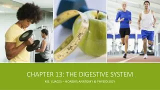
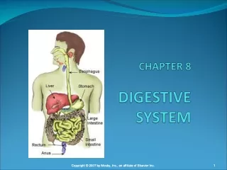
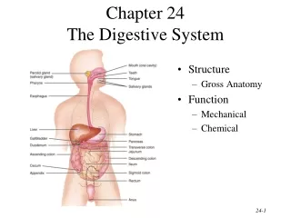
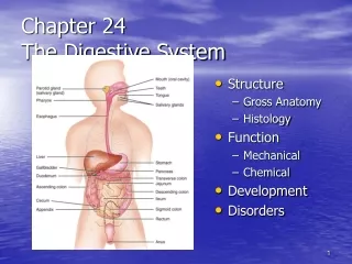
Chapter 17 - Digestive System. Digestion - process by which food is changed into forms that can be absorbed through cell membranes. Major Organs. General Characteristics of the Alimentary Canal (GI Tract). Extends from mouth to anus (about 9m long)

E N D
Chapter 17 - Digestive System • Digestion - process by which food is changed into forms that can be absorbed through cell membranes.
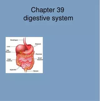
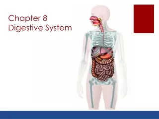
General Characteristics of the Alimentary Canal (GI Tract) • Extends from mouth to anus (about 9m long) • Organs include: mouth, pharynx, esophagus, stomach, small intestine, and large intestine • Accessory organs: salivary glands, liver, gall bladder, pancreas
Structure of the wall- 4 layers • 1. Mucosa • 2.Submucosa • 3. Muscularis • 4. Serosa
Mucosa • innermost layer • made of epithelial and connective tissue and some smooth muscle • has glands that secrete mucus for lubrication and protection from digestive enzymes
Submucosa • beneath mucosa • contains connective tissue, glands, blood vessels lymph vessels, and nerves • nourishes mucosa and carries absorbed nutrients away
Muscularis • 2 layers of muscle (smooth) • circular muscle layer around submucosa • longitudinal layer around circular layer • function is to move food through the canal (mixing & peristalsis
Serosa • outermost layer • protects underlying tissues • secretes serous fluid to lubricate outer tube so organs slide freely against each other
. Movements of the tube • 1. Mixing • Smooth muscles contract rhythmically • Mix food, digestive juices, and mucus • 2. Peristalsis • Wavelike motion of longitudinal muscle layer to move food along • Begins when food expands the tube
Movements of the Tube • mixing movements • peristalsis
Innervention of the Tube 1. Parasympathetic NS (autonomic NS) • Increases digestive activities • In control under normal, restful conditions 2. Sympathetic NS (autonomic NS) • In control in stressful situations • Contract sphincter muscles (blocks movement of food) - found between organs of GI tract • Inhibits digestive activities
Mouth (Fig 17.7 page 650) • Receives food and starts digestion by chewing and mixing with saliva • Cheeks and lips Tongue • Muscular organ that mixes and moves food
Palate • roof of mouth • Hard palate - anterior portion • Soft palate - posterior portion • Uvula - extension of soft palate • Tonsils - masses of lymphatic tissue - Palatine tonsils (lateral to palate) - Pharyngeal tonsils = adenoids (posterior pharynx)
Mouth • ingestion • mechanical digestion • prepares food for chemical digestion
Palate • roof of oral cavity
Teeth • 2 sets develop in sockets in mandibular and maxillary bones • 20 primary (deciduous) lost between 6-12 years • 32 secondary (permanent) • function is to break food into smaller pieces
Primary Teeth • 8 incisors • 4 cuspids • 8 bicuspids • 12 molars
4 types of teeth - incisors, cuspid, bicuspids, molars (fig 17.9 page 656)
tooth structure (fig 1 7.10 page 657) • crown = exposed area of tooth • root = area below gum (gingiva) • enamel = covering on crown made ofCa+ salts (hardest substance in body) • dentin = bulk of tooth under enamel
pulp = central cavity that contains blood vessels, nerves, and connective tissue • cementum = encloses root • periodontal ligament = attaches tooth to jaw • See Clinical application 17.1 page 657 - effects of bacteria on teeth
Salivary Glands • Secretes saliva to moisten and bind food and begins digestion of carbohydrates • Helps cleanse mouth and regulate pH (6.5 - 7.5) • Makes taste possible
Salivary secretions • Serous cells secrete amylase (enzyme) to start carbohydrate digestion • Mucus cells secrete mucus for lubrication
Major Salivary Glands (fig 17.11 page 658) Parotid • Anterior to ear • largest • secrete saliva rich in amylase Submandibular • located on floor of mouth • secrete viscous saliva
Sublingual • located inferior to tongue • secretes mostly mucus
Pharynx (throat) • divided into 3 parts Nasopharynx • superior to soft palate • passageway for air during breathing Oropharynx • posterior to mouth • passageway for food and air moving to and from nasal cavity
Laryngopharynx • inferior to oropharynx • passageway to esophagus
Esophagus • Straight collapsible tube about 25cm long • Passageway for food from pharynx to stomach • Posterior to trachea (tube to lungs) • See hiatal hernia page 661 • Lower esophageal sphincter muscles usually remains contracted to prevent regurgitation of stomach contents
Stomach • J shaped pouchlike organ 25-30cm long • Inner has thick mucosal folds called rugae - pg 662 fig 17.17 • Pyloric sphincter muscle found at entrance to small intestine (duodenum) • Mixes food with gastric juice, initiates digestion of proteins, carries on a limited amount of absorption, and moves food to small intestines.
Gastric secretions • Mucosa of stomach has gastric pits that are the openings of gastric glands. Page 664 fig 17.19 • Gastric juice contains: see page 665 table 17.5
Gastric Secretions • mucus • from goblet cells and mucous glands • protective to stomach wall • pepsinogen • from chief cells • inactive form of pepsin • pepsin • from pepsinogen in presence of HCl • protein splitting enzyme • intrinsic factor • from parietal cells • required for vitamin B12 absorption • hydrochloric acid • from parietal cells • needed to convert pepsinogen to pepsin
Regulation of Gastric Secretions Parasympathetic NS (normal conditions) - stimulates release of the hormone gastrin which increases the activity of gastric glands - hormone somatostatin inhibits acid secretion
Gastric Absorption • Stomach wall not well adapted to absorb digestive products • Only small quantities of water, certain salts, alcohol and some lipid soluble drugs are absorbed
Mixing and emptying actions • Mixing of food + gastric juice = chyme • Peristalsis moves chyme toward small intestine • Pyloric sphincter relaxes (opens) & chyme is pushed a little at a time into small intestine • Enterogastric reflex inhibits peristalsis in stomach as small intestine fills
Enterogastric Reflex regulates the rate at which chyme leaves the stomach
Vomiting • Caused by irritation or distension in stomach or intestines • Can be stimulated by drugs, toxins in foods, rapid changes in body motion, sights, sounds, odors, tastes, emotions and mechanical stimulation of back of pharynx • Vomit center is in medulla oblongata • Stomach is squeezed from all sides forcing contents upward • Nausea caused by activity in or near vomit center
Pancreas (accessory organ) • Secretes digestive juice called pancreatic juice Structure • Its head is found in C-shaped curve of duodenum and tail against the spleen • Page 668 Fig 17.23 • Pancreatic duct connects to small intestine at the same place as the bile duct from the liver and gallbladder