Download

1 / 23
230 likes | 351 Views
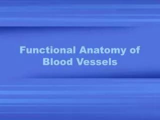
Biology 161 Lab 4 - Blood Vessels, Lymphatics , Pressure Points, Surface Anatomy. Scott.lehbauer@lethbridgecollege.ab.ca. The Arteries. The Circle of Willis.

E N D
Biology 161 Lab 4 - Blood Vessels, Lymphatics, Pressure Points, Surface Anatomy Scott.lehbauer@lethbridgecollege.ab.ca
The Arteries The Circle of Willis Circle of Willis - also called the cerebral arterial circle this structure surrounds the pituitary gland and optic chiasma. It connects the anterior and posterior blood supply to the brain. It also equalizes blood pressure in the two brain regions and provides alternate routes for blood to reach the brain tissue in case of blockage to the carotid or vertebral arteries.
The Circle of Willis Basilar Artery Internal Carotid Artery Vertebral Arteries
Arteries of the Neck and Face Internal Carotid Artery External Carotid Artery Superficial Temporal Artery Common Carotid Artery Vertebral Artery
Arteries coming off the Heart Right Common Carotid Left Common Carotid Right Subclavian Right Brachiocephalic Left Subclavian Aorta
Arteries of the Arm Axillary Artery Circumflex Scapular Artery
Arteries of the Arm Cont. Brachial Artery Radial Artery Median Artery Ulnar Artery Superficial Palmer Arch
Coronary Arteries (Left Side) Aortic Arch Left Coronary Artery Anterior Descending Artery Circumflex Artery
Right Coronary Artery and Cardiac Vein Great Cardiac Vein Right Coronary Artery
Arteries of the Torso Gastric Artery Stomach
Arteries of the Torso (Superior Mesenteric) Superior Mesenteric Artery Superior Mesenteric Artery
Arteries of the Torso (Inferior Mesenteric) Inferior Mesenteric Artery Inferior Mesenteric Artery
Arteries of the Torso Hepatic Artery Liver
Arteries of the Torso (Celiac Trunk) Celiac Trunk Splenic Artery
Arteries of the Torso (Kidneys) Renal Artery Renal Vein
Arteries of the Torso (Iliac Arteries) Common Iliac Artery External Iliac Artery Internal Iliac Artery
Arteries of the Leg Femoral Artery
Arteries of the Leg Anterior Tibial Artery Popliteal Artery
Pressure Points Superficial Temporal a. - Are arteries that when compressed can stop blood flow into distal tissues during hemorrhage. Facial a. Subclavian a. Carotid a. Brachial a. Radial a. Femoral a. Popliteal a. Dorsal Pedal a.
Lymphatic System • Lymph – Protein containing fluid transported by lymphatic vessels. • Lymphatics – an elaborate system of drainage vessels that collect the excess protein-containing interstitial fluid and return it to the bloodstream. • Lymph Node – a small lyphoid organ that filters lymph; they contain macrophages and lymphocytes.
Lymphatic System Cervical Thoracic Axillary Abdominal Cubital Inguinal Pelvic Popliteal
Lymphatic System Inguinal Lymph Nodes Axillary Lymph Nodes
Spirometry Tidal Volume – The amount of air that moves into then out of the lungs during normal quiet breathing. Vital Capacity – The total amount of exchangeable air in the lungs. Or the total amount of air blown out during one forced exhalation. There are 3 factors which influence vital capacity: 1.) Age 2.) Sex 3.) height