Download

1 / 15
170 likes | 395 Views
Objectives Discuss the anatomical structure of the trachea with its relations. Define the term bronchial tree. Describe bronchopulmonary segments. The Trachea. The trachea is a mobile cartilaginous and membranous tube. It begins below the cricoid cartilage at C6 vertebra .

E N D
Objectives Discuss the anatomical structure of the trachea with its relations. Define the term bronchial tree. Describe bronchopulmonary segments.
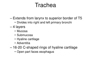
The Trachea • The trachea is a mobile cartilaginous and membranous tube. • It begins below the cricoid cartilage at C6 vertebra. • It descends in the midline of the neck. • Ends in the thorax by dividing into right and leftprincipal (main) bronchi at the level of the sternal angle (opposite the disc between the fourth and fifth thoracic vertebrae).
In adults the trachea is about 4آ½ in. (11.25 cm) long and 1 in. (2.5 cm) in diameter. • It is kept patent by the presence of U-shaped bars (rings) of hyaline cartilage which are connected by smooth muscle, the trachealis muscle.
Relations of the Trachea in the Neck: • Anteriorly: Skin, fascia, isthmus of the thyroid gland (in front of the second, third, and fourth rings), inferior thyroid vein. • Posteriorly: Right and left recurrent laryngeal nerves and the esophagus • Laterally: Lobes of the thyroid gland and the carotid sheath.
The relations of the trachea in the superior mediastinum • Anteriorly: The sternum, the thymus, the left brachiocephalic vein, the origins of the brachiocephalic and left common carotid arteries, and the arch of the aorta . • Posteriorly: The esophagus and the left recurrent laryngeal nerve . • Right side: The azygos vein, the right vagus nerve, and the pleura . • Left side: The arch of the aorta, the left common carotid and left subclavian arteries, the left vagus and left phrenic nerves, and the pleura .
Blood Supply of the Trachea: • The upper two thirds are supplied by the inferior thyroid arteries and the lower third is supplied by the bronchial arteries. • Lymph Drainage: • The lymph drains into the pretracheal and paratracheal lymph nodes and the deep cervical nodes. • Nerve Supply: • The sensory nerve supply is from the vagi and the recurrent laryngeal nerves. Sympathetic nerves supply the trachealis muscle.
Bronchial tree means the bronchi and their branching structures. • The trachea bifurcates behind the arch of the aorta into the right and left principal (primary, or main) bronchi . • The right principal (main) bronchus is wider, shorter, and more vertical than the left & about 1 in. (2.5 cm) long . • It divides into a superior, middle, and an inferior lobar bronchus.
The left principal (main) bronchus is narrower, longer, and more horizontal than the right and is about 2 in. (5 cm) long. • It divides into a superior and an inferior lobar bronchus.
The bronchi divide dichotomously, giving rise to several million terminal bronchioles. • Terminal bronchioles terminate in one or more respiratory bronchioles. • Each respiratory bronchiole divides into 2 to 11 alveolar ducts that enter the alveolar sacs. • The alveolar sacs form alveoli.