Download
1 / 1
10 likes | 190 Views
Opacidades lineales. Opacidades nodulares. Aumento de la densidad radiológica. Disminución de la densidad radiológica. Localización anomalías (lobulillo pulmonar secundario). Distribución anomalías. CAPACIDAD PREDICTIVA PREVALENCIA HALLAZGO AP.

E N D
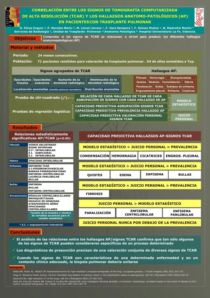
Opacidades lineales Opacidades nodulares Aumento de la densidad radiológica Disminución de la densidad radiológica Localización anomalías (lobulillo pulmonar secundario) Distribución anomalías CAPACIDAD PREDICTIVA PREVALENCIA HALLAZGO AP CAPACIDAD PREDICTIVA VALORACIÓN PERSONAL SIGNOS TCAR Fibrosis Hemorragia Bronquiectasias Quistes Nódulos Consolidación Edema Panalización Bullas Subtipos de enfisema Engrosamiento pleural Enfisema Cicatrices CAPACIDAD PREDICTIVA HALLAZGOS AP-SIGNOS TCAR MODELO ESTADÍSTICO = JUICIO PERSONAL = PREVALENCIA CONDENSACIÓN HEMORRAGIA CICATRICES ENGROS. PLEURAL MODELO ESTADÍSTICO > JUICIO PERSONAL > PREVALENCIA ENFISEMA BULLAS QUISTES EDEMA MODELO ESTADÍSTICO = JUICIO PERSONAL > PREVALENCIA FIBROSIS ENFISEMA CENTRILOBULAR ENFISEMA PANLOBULAR PANALIZACIÓN JUICIO PERSONAL > MODELO ESTADÍSTICO JUICIO PERSONAL NUNCA POR DEBAJO DE LA PREVALENCIA CORRELACIÓN ENTRE LOS SIGNOS DE TOMOGRAFÍA COMPUTARIZADA DE ALTA RESOLUCIÓN (TCAR) Y LOS HALLAZGOS ANATOMO-PATOLÓGICOS (AP) EN PACIENTESCON TRASPLANTE PULMONAR D. Pérez Enguix 1, P. Morales Marín 2, M. Lloret Llorens 1, F. Vera Sempere 3, F. Gómez Muñoz 1, N. Madroñal Martín 1 Servicios de Radiología 1, Unidad de Trasplante Pulmonar 2.Anatomía Patológica 3. Hospital Universitario La Fe. Valencia. Comprobar si los signos de TCAR se relacionan, o sirven para predecir, los diferentes hallazgos anatomopatológicos (AP) Objetivos Material y métodos Periodo: 24 meses consecutivos. Población: 71 pacientes remitidos para valoración de trasplante pulmonar . 54 de ellos sometidos a Txp. Signos agrupados de TCAR Hallazgos AP: RELACIÓN DE CADA HALLAZGO DE TCAR DE CADA AGRUPACIÓN DE SIGNOS CON CADA HALLAZGO DE AP Prueba de chi-cuadrado (c2) : MODELO ESTADÍSTICO CAPACIDAD PREDICTIVA AGRUPACIÓN SIGNOS TCAR Pruebas de regresión logística: JUICIO PERSONAL Resultados Relaciones estadísticamente significativas AP/TCAR (p<0.05) Fibrosis VIDRIO DELUSTRADO SIGNO INTERFASE E.I*. INTRALOBULAR E.I. SUBPLEURAL E.I. INTERLOBULAR Edema OPACIDAD INTERLOBULAR Enfisema ENFISEMA TCAR E.I.PERIBRONCOVASCULAR BANDAS PARENQUIMATOSAS ENFISEMA CENTRILOBULAR LUCENCIA PANLOBULAR BULLAS Bullas ENFISEMA BULLAS ENFISEMA CENTROLOBULAR Bronquiectasias NÓDULOS CENTRILOBULILLARES BRONQUIECTASIAS MOSAICO DE DENSIDAD ATRAPAMIENTO AÉREO OPACIDADES CENTRILOBULILLARES Tamaño de la muestra y número de variables excesivas para el modelo estadístico * E.I. = engrosamiento intersticial Conclusiones • El análisis de las relaciones entre los hallazgos AP/signos TCAR confirma que tan sólo algunos de los signos de TCAR pueden considerarse específicos de un proceso determinado • Los diagnósticos de presunción precisan de una valoración conjunta de diversos signos de TCAR • Cuando los signos de TCAR son característicos de una determinada enfermedad y en un contexto clínico adecuado, la biopsia pulmonar debería evitarse Bibliografía Webb WR, Muller NL, Naidich DP. Standardized terms for high-resolution computed tomography of the lung: a proposed glossary. J Thorac Imaging 1993; 8(3):167-175 Hogg JC. Benjamin Felson lecture. Chronic interstitial lung disease of unknown cause: a new classification based on pathogenesis. AJR Am J Roentgenol 1991;156(2):225-33 Kazerooni EA. High-resolution CT of the lungs. AJR Am J Roentgenol 2001;177:501-519. Wells AU, King AD, Rubens MB, Cramer D, du Bois RM, Hansell DM. Lone cryptogenic fibrosing alveolitis: a functional -morphologic correlation based on the extent of disease on thin-section computed tomography. Am J Respir Crit Care 1997;155:1367-75.