Download
1 / 30
340 likes | 1.07k Views
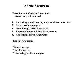
Pseudohypertension Osler’s Sign and Aortic Arch Calcification. Case Report. OB – 89 y/o man admitted with SOB, cough, knee pain Past Medical History Hypertension Mild CCF Prostate Ca Chronic renal impairment (Cr. 250) Medications ACE I Amlodipine

E N D
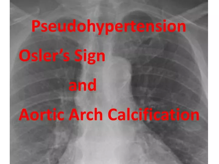
Pseudohypertension Osler’s Sign and Aortic Arch Calcification
Case Report OB – 89 y/o man admitted with SOB, cough, knee pain Past Medical History • Hypertension • Mild CCF • Prostate Ca • Chronic renal impairment (Cr. 250) • Medications • ACE I Amlodipine • Aspirin Fruesemide 40mg
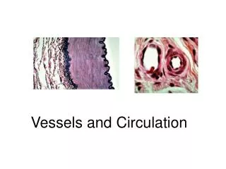
Pseudohypertension • When BP measured by cuff is falsely elevated compared to reference standard because of hardened calcific arterial walls • Pathophysiology - arterial calcification as opposed to atherosclerosis/collagen deposition* • Associations • Age - Hypertension • Atherosclerosis - Scleroderma • Prevalence - 1.7% and 2.5% but poorly studied* * Zuschke et al, Pseudohypertension, Southern Medical Journal 1995, 88:1185-90
Atherosclerotic calcification: • Intimal layer – cellular necrosis, inflammation and lipid deposition • As lesion progresses, osteogenesis • Typical vascular risk factors, especially DM and smoking Monckeberg’s Sclerosis (medial artery calcification) • Age, diabetes and renal disease • Related to PTH, calcium and phosphate product, vitamin D and uraemia • Bone-associated cells/proteins important
Osler’s Maneuver “It may be difficult to estimate how much of the hardness and firmness is due to the tension of blood within the vessel and how much to the thickening of the wall. For example, when the radial artery can be felt beyond the point of compression, it’s walls are sclerosed” Sir William Osler, 1892
Osler’s Maneuver • Term coined by Messerli et al (N Engl J Med) in 1985: • Oslers, sphygmo, ECHO • Strong association between Osler’s and PsHTN • Usefullness refuted by Prochazka et al (Clin Res) in 1987, due to poor inter-observer reliability • Subsequently, interobserver agreement 82% with no training effect# • Prevalence • 1.7%* to 12.3%** (with 0% under 50y/o and 15.6% in over 65y/o)*** • Prevalence increases with age, history of hypertension or stroke** # Hla et al, Observer vasriability of oslers maneuver in detection of pseudohypertension, J Clin Epi 1991, 44:513-18 * Kuwajima et al, Pseudohypertension in the elderly, J. Hyperten, 1990, 8:429-32 ** Wright and Looney, Prevalence of positive osler’s manouvre in 3397 persons screened for the SHEP, Journal of Human Hypertension, 1997, 11:285-289 ***Prochazka et al, Oslers maneuver in outpatient veterans, J Clin Hypertension, 1987, 3:554-8
Osler’s Maneuver – previous studies Systematic review using MEsH terms pseudohypertension, Osler’ssign and Osler’smaneuver revealed 8 studies: • Two examining prevalence of Osler’s sign • One examining observer variability in Osler maneuver • Four comparing radial artery to sphyg in osler-positive • One comparing radial doppler to sphyg in osler-positive
Aortic Arch Calcification • Thoracic aortic calcification has been associated with increased cardiovascular mortality (hazard ratio between 3 - 6 for IHD and 2.3 for CerebroVD mortality)* • Strong association with increasing age, hypertension, pulse pressure and smoking • less association with other known cardiovasc risk factors and CRP** (these studies DID NOT include renal function) • Clear evidence of association with renal failure • Vitamin D, PTH, calcium and phosphate, ureamia • Genetic component*** • * Jacobs et al, Comparing coronary artery calcium and thoracic aortic calcium for prediction of all-cause mortality and cardiovascular events in low-dose non-gated computed tomography in a high-risk setting of heavy smokers, Atherosclerosis, 2010, 209:455-62 • * Calcification of the thoracic aorta as detected by spiral computed tomography among stable angina pectoris patients, Circulation, 2008, 118:1328-34 • ** Takasu et al, Relationship of thoracic aortic wall calcification to cardiovascular risk factors: the multi-ethnic study of atherosclerosis (MESA), American Heart Journal, 2008, 155(4) • *** Parikh et al, Parental occurrence of premature cardiovascular disease predicts increased coronary artery and abdominal aortic calcification in the framingham offspring and third generation cohorts, Circulation, 2007, 116:1473-81
Oslers maneuver, Pseudohypertension and Aortic Arch Calcification Simon Quilty Nick Collins Nick Jackson Paul Puller Angela Puller John Attia
Study Design • Sequential patients undergoing non-emergency cardiac catheterization in RNC Cath Lab • Verbal consent – 100% acceptance • Study participants underwent: • Pre-procedure questionnaire • If recent CXR in past 5 years, Aortic Arch Calcification score calculated • Pre-procedural manual and automatic sphygmo BP • Study blood pressure measurements Peripheral transduced BP Automatic sphygmo BP (on non-procedure arm) Central transduced BP
Questionnaire • Age, sex • diabetes • hypertension • hyperlipidaemia • Past or current smoking • Number of pack years smoked • Past history ischaemic heart disease • Past history stroke • Past history peripheral vascular disease
Aortic arch calcification calculation Tetsuya et al, Simple evaluation of aortic arch calcification by chest radiography in haemodialysis patients, Hemodialysis international, 2009, 13:301-306
Statistical Analysis • Inter-rater reliability of Osler’s Sign • Kappa = 0.54 • Inter-observer agreement = 89%
Pearson’s correlation - Sphygmo vs Peripheral Transduced BP No correlation between systolic Correlation between diastolic, P < 0.0001, R = 0.55 Systolic Diastolic Difference auto vs transduced Automatic Sphyg BP Automatic Sphyg BP
Sphygmo vs Central TransducedBP No correlation between systolic Correlation between diastolic, P < 0.0001, R = 0.49 Systolic Diastolic Difference auto vs transduced Automatic Sphygmo BP Automatic Sphygmo BP
Osler’s Sign and Pseudohypertension • Fisher’s exact test Pseudohypertension as defined as >10mmHg over-estimate of reference (transduced) BP No statistically significant association between Osler’s Sign and defined pseudohypertension centrally or peripherally, systolic or diastolic
Osler’s Maneuver and Pseudohypertension • Unpaired t-test • Osler’s Sign and magnitude of difference between automatic and transduced BP: • Systolic Pseudohypertension no statistically significant difference • Diastolic pseudohypertension statistically significant when measured centrally or peripherally • Central – 4mmHg between osler’s pos/neg (P<0.03) • Peripheral – 16mmHg between osler’s pos/neg (P<0.0001) • Patients with a positive osler’s maneuver had a diastolic cuff pressure that was on average 16mmHg above transduced
Osler’s Sign and Aortic Arch Calcification • There was a statistically significant correlation between a positive and negative osler’s sign and extent of aortic arch calcification: • Osler’s positive – mean AoAC score = 6.7 • Osler’s negative – mean AoAC score = 3.28 • P = 0.004
Stepwise Multiple Linear Regression – Magnitude of difference in BP • Oslers sign +/- • Age • Sex • Aortic arch calcification score (0-16) • eGFR • Previous or current smoker • Number of pack yrs of smoking • History of Diabetes • History of HTN • History of dyslipidaemia • Previous history of IHD • Previous history of Stroke • Previous history of PVD
Magnitude of difference in BP - Automatic vs Peripheral transduced • Systolic • Stroke (-9.77mmHg if +ve Hx, P = 0.01) • Diastolic • Osler’s Sign positive (+5.28mmHg if Oslers +ve, P = 0.07) • History of IHD (-3.70mmHg if +ve Hx, P = 0.07)
Stepwise Multiple Linear Regression Aortic Arch Calcification • Same variables plus pressure difference between sphygmo and transducer (systolic, diastolic, central and peripheral) • Age (+0.08 per yr of age, P = 0.03) • Renal function (-0.06 per eGFR, P = 0.008) • Osler’s sign (+2.32 if Oslers +ve, P = 0.07)
Conclusions • Pseudohypertension leads to over-treatment of blood pressure • There is no “gold standard” blood pressure • In high-risk patients with resistant diastolic hypertension, an Osler’s Maneuver may be useful • Aortic arch calcification does not assist in risk stratifying in regards to pseudohypertension
Conclusions Measurement of BP is imprecise however there are strategies that improve accuracy of diagnosis* *Powers et al, Measuring blood pressure for decision making and quality reporting: where and how many measures? Annals of Internal Mericine, 2011, 154:781-88
Within patient SBP variance and number of measurements *Powers et al, Measuring blood pressure for decision making and quality reporting: where and how many measures? Annals of Internal Medicine, 2011, 154:781-88
Concurrence between automatic and manual sphygmomanometer Linear regression, Two-sided P <0.0001, R = -0.32 (sys) R = 0.06 (dias) Systolic Diastolic Difference auto vs manual Automatic BP Automatic BP
Central transduced vs Peripheral transduced BP Close to statistically significant correlation for systolic (P = 0.051, R=0.24) Statistically significant correlation for diastolic (P < 0.0001, R=0.48 ) Systolic Diastolic Difference central vs peripheral Central BP Central BP