Download
1 / 14
220 likes | 485 Views
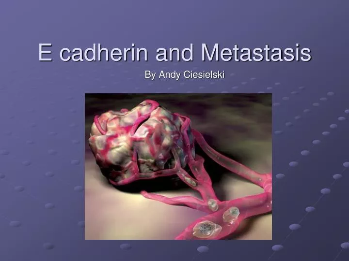
E cadherin and Metastasis. By Andy Ciesielski. CDH1 gene. Encodes E Cadherin 16q22.1 Ca2+ Dependent Cell-Cell Adhesion Molecule 16 exons.

E N D
E cadherin and Metastasis By Andy Ciesielski
CDH1 gene • Encodes E Cadherin • 16q22.1 • Ca2+ Dependent Cell-Cell Adhesion Molecule • 16 exons Genomic organization of the human E-cadherin gene. Positions of exons are shown in color boxes with the base pair number of each exon. The connecting lines are introns.
5 Extracellular Domains containing conserved repeated Amino Acid sequences (cadherin repeats),1 Transmembrane Domain, 1 Intracellular Domain • Forms Homophilic Interactions with Adjacent E cadherins in Lateral Dimerization • Dynamically Associates with Alpha, Beta, and Gamma Catenins and p120 • 120 kDa Shematic illustration of E-cadherin in adherens junction. E-cadherin homodimer on the cytoplasmic membranes of adjacent cells is shown. The juxtamembrane region with the interacting molecules is also shown. CM – cytoplasmic membrane; AJ – adherens junction; ED – extracellular domain; ID – intracellular domain; AC – actin cytoskeleton; 1-beta-catenin; 2-alpha-catenin; 3-p120.
Cellular Roles • Adhesion Between Epithelial Cells- Adherens Junctions • Development- Gastrulation, Neurulation, Organogenesis • Adhesion of blastomeres and embryo compaction • Controlled epithelial mesenchymal conversion (loss of epithelial adhesion and polarity) • -/- mutations lethal at blastocyst stage in mice
Cellular Roles • Signaling Pathways • EGFR and MAPK • Wnt • Snail, Slug, Twist- Repressors of E cadherin • Tumor Suppressor
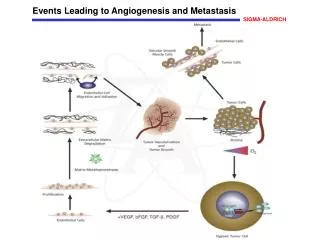
Metastasis • Tumor cells lose adhesion for surrounding cells, undergoing EMT • Spread into blood vessels and other tissues forming new tumors at new sites
Loss of E cadherin Leads to Metastatic Tumors • E cadherin functions as a Tumor Suppressor • E cadherin loss enables disaggregation of cancer cells from one another • LOH in CDH1 Correlates with metastasizing malignancies • Over 75% of Metastatic Cancers Exhibit CDH1 Abnormalities • CDH1 mutations involved with several cancers: Breast, Liver, Prostate, Stomach, Endometrium, Ovary, and Lung • Loss of E cadherin Function Correlates with Poor Prognosis
E Cadherin Expression in Cancer • Type 1- Preserved Type, Tumor Cells Retain Cadherin Expression • Type 2- Reduced Type, Tumor Cells Show Reduced Cadherin Expression, • Type 3- Complete Loss of Cadherin Function • Type 4- Aggregate Failure
E Cadherin Mutations • Hypermethylation • Somatic Mutations- Insertions, Deletions, Non-sense Mutations • Diffuse Gastric Tumor Showed In-Frame Mutations • Infiltrative Breast Cancers Showed Out-of-frame Mutations Leading to Truncated E Cadherins • Hereditary Predispositions • Early onset cancers, high penetrance • Silencing or Over Activation of Various Influential Factors • CDH1 promoter • Snail, Slug, Twist – trigger EMT, repress E cadherin • Tyrosine Phosphorylation of accessory proteins, Alpha, Beta, Gamma Catenins interfering with Cadherin Action
Down Regulation of E cadherin and Dominant Negative E cadherin Proteins Results in Loss of Typical Morphology- Loss of Cellular Adhesion The adoption of fibroblastic morphology by shEcad cells typifies a EMT shEcad cells also showed increases in Mesenchymal proteins N-Cadherin and Vimentin compared to control cells
Orthotopic and Tail Vein Assays Reveal Metastases in Lungs of Nude Mice shCntrl shEcad DN-Ecad
Motility and Apoptosis of Cells Upon Loss of Cell Adhesion shEcad cells showed increased motility and invasiveness in Boyden Chamber Assays shEcad cells showed high levels of cells indicating decreased apoptosis in the absence of substrate attachment shEcad cells showed decreased amounts of annexinV indicating lower rates of apoptosis
Summary • E cadherin normally functions to maintain cell adherens junctions in epithelial tissues • E cadherin can be appropriately regulated to trigger EMT during development • Inappropriate regulation or mutations is strongly correlated with a variety of metastatic cancers • E cadherin loss appears sufficient to give metastatic ability HMLER cells. • Loss of extracellular and intracellular domains is necessary for gaining metastatic ability • Loss of E cadherin with additional oncogenic lesions quickens the transition to invasive metastases
References • Takeichi, Masatoshi, Cadherins in Cancer: Implications for Invasion and Metastasis. Cell Biology 5 (1993) 806-811. • T. Onder, P. Gupta, S. Mani, J. Yang, E. Lander, R. Weinberg. Loss of E-Cadherin Promotes Metastasis via Multiple Downstream Transcriptional Pathways. Cancer Research 68 (2008) 3645-3654. • Pecina-Slaus, Nives. Tumor Repressor Gene E-Cadherin and its Role in Normal and Malignant Cells. Cancer Cell International 3 (2003) 1-7.