Download
1 / 74
850 likes | 1.17k Views
DR.T.RAMANI DEVI MD DGO FICS FICOG CONSULTANT OBSTETRICIAN AND GYNAECOLOGIST RAMAKRISHNA NURSING HOME, TRICHY. M ullerian anomalies. INTRODUCTION. MDA are fascinating disorders to obstetricians and gynaecologists MD forms tubes, uterus, cervix and upper part of vagina

E N D
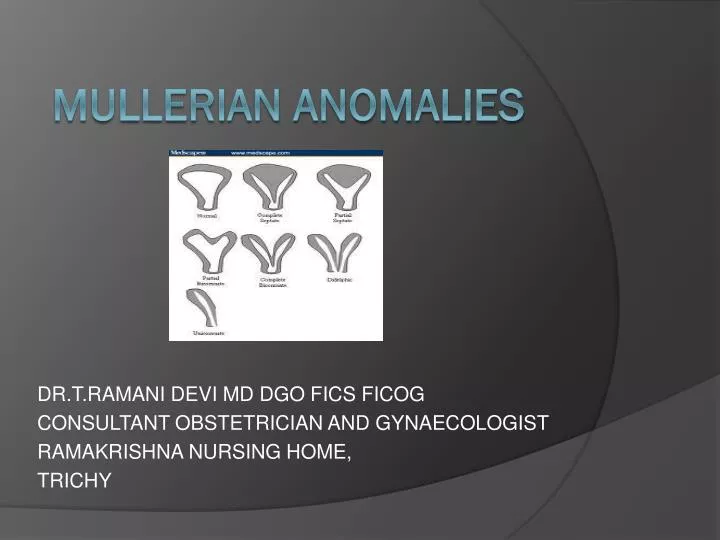
DR.T.RAMANI DEVI MD DGO FICS FICOG CONSULTANT OBSTETRICIAN AND GYNAECOLOGIST RAMAKRISHNA NURSING HOME, TRICHY Mullerian anomalies
INTRODUCTION • MDA are fascinating disorders to obstetricians and gynaecologists • MD forms tubes, uterus, cervix and upper part of vagina • Ranges from agenesis to duplication. • Associated with renal and axial skeletal systems anomalies • Has varying presentation ranging from primary amenorrhea to menstrual disorders, infertility and pregnancy complications like BOH, PTL, Ectopic , etc • MDA has varying treatment from ability to have coitus to conceive and deliver normal babies.
INCIDENCE • Dates back to 16th century a case utero vaginal agenesis – Columbo et al (1600) • General population – 0.1-3.5% - Byrene et al • Fertile women – 4.3% • Infertile women – 3.6% • Sterile group - 2.4% • Recurrent Aborters 5 - 13% - Grimbizis et al
ETIOLOGY • Dysregulation occuring in differentiation, migration, fusion and canalisation • Associated with renal anomalies, axial skeletal anomalies and rarely cardiac and auditory anomalies • Probable causes: Intrauterine infection , genetic aberration, Teratogens like DES and Thalidomide.
GENETICS OF MDA • Sporadic • Familial • Multifactorial • Autosomal dominant • Autosomal recessive • X linked • Variants of GALT (Galactose 1 phosphate uridyltransferase enzyme defect) Genes Associated :- HOXA 9, 13 & WNT 4
CLASSIFICATION OF MDA • 1979 – Buttram and Gibbons classification Modified • 1988 – American Fertility Society classification
American Fertility Society Classification of Mullerian Anomalies
INCIDENCE OF MDA ACCORDING TO AFS • Arcuate uterus 32.8% • Septate uterus 33.6% • Bicornuate uterus 20.0% • DES exposed uterus 0.8% • Unicornuate • Uterine didelphys 33%
EFFECT OF MDA UPON REPRODUCTION • Infertility • Endometriosis • Ectopic pregnancy • Recurrent Pregnancy Loss • Prematurity , IUGR , fetal malposition • Uterine dysfunction • Uterine rupture • Increased perinatal morbidity and mortality
DIAGNOSIS OF MDA • Clinical • Hystero salphingogram • Sonosalphingogram • MRI – 100% accuracy • Hystero laparoscopy • Laparotomy or LSCS
Vulvar Abnormalities Vulval and lower 1/3rd vagina atresia Labial Fusion • Most commonly due to congenital adrenal hyperplasia. Imperforate hymen • Persistence of the fusion between the sinovaginal bulbs at the vestibule • Associated with primary amenorrhea and hematocolpos
Vaginal Abnormalities Developmental abnormalities of the normal single vagina include: Vaginal agenesis Vaginal atresia Double vagina Longitudinal vaginal septum Transverse vaginal septum
Obstetrical significance of vaginal abnormalities • Complete mullerian agenesis – pregnancy is impossible because uterus and vagina is absent • About one third of women with vaginal atresia have associated urological abnormalities • Complete vaginal atresia– precludes intercourse and then pregnancy • In most cases of partial atresia, because of pregnancy-induced tissue softening, obstruction during labor is gradually overcome. interferes with descent
Obstetrical significance of vaginal abnormalities Complete longitudinal vaginal septumusually does not cause dystocia because half of the vagina through which the fetus descends dilates satisfactorily. Incomplete septum, however, occasionally interferes with descent.
Cervical Abnormalities Atresia. This may be combined with incomplete development of the upper vagina or lower uterus Double cervix. Each distinct cervix results from separate müllerian duct maturation. Both septate and true double cervices are frequently associated with a longitudinal vaginal septum. Many septate cervices are erroneously classified as double. Single hemicervix. This arises from unilateral müllerian maturation. Septate cervix. This consists of a single muscular ring partitioned by a septum. The septum may be confined to the cervix, or more often, it may be the downward continuation of a uterine septum or the upward extension of a vaginal septum.
CLASS I- ROKITANSTY SYNDROME • Primary amenorrhea • Feminine patients • Short vagina DD: Testicular feminization syndrome
INVESTIGATIONS • Karyotyping • USG/MRI • Hormone assay • IVP (associated vertebral anomalies can be detected) and renal sonography • Diagnostic Laparoscopy is not routinely done.
TREATMENT • Vaginal Reconstruction – Vagino plasty : Mac Indoes Vaginoplasty; Williams vulvovaginoplasty, Vecchietti procedure • Fertility – by surrogacy • Psychological support
Unicornuate Uterus (Class II) • Women with a unicornuate uterus have an increased incidence of infertility, endometriosis, and dysmenorrhea. • Implantation in the normal-sized hemiuterus is associated with increased incidence of: • spontaneous abortion • preterm delivery • intrauterine fetal demise
UNICORNUATE UTERUS • Unilateral failure of development of MDA Incidence: 2.5-13% Types : Unicornuate Unicornuate with rudimentary horn -Communicating -Non communicating - with endometrium -without endometrium Associated Renal anomalies like renal agenesis, Horseshoe kidney and pelvic kidney44% (In the presence of obstructed horn)
CLINICAL FEATURES • Haematometra • Endometriosis • Preterm labour – 43% • IUGR • Mal presentation • Ectopic -4.3% • Pregnancy in accessory horn -2% • Rupture uterus
IMAGING MODALITIES IN UNICORNUATE UTERUS HSG 3D USG MRI
DIAGNOSIS AND SURGICAL MANAGEMENT • HSG – non communicating horn cannot be diagnosed • USG – 3D or High Resolution • MRI – banana shaped uterus • Laparoscopy – indicated for excision of rudimentary horn which has endometrium • IVU or renal sonography Cervical encirclage is mandatory if patient conceives
REPRODUCTIVE OUTCOME IN UNICORNUATE UTERUS • Live birthrate 43.7% • Abortion rate 35-43% • Preterm delivery 27% • Term delivery 31%
NONCOMMUNICATING RUDIMENTARY UTERINE HORN * attached fallopian tube (arrow) was patent*
Uterine Didelphys (Class III) • This anomaly is distinguished from bicornuate and septate uteri by the presence of complete nonfusion of the cervix and hemiuterine cavity • Except for ectopic and rudimentary horn pregnancies, problems associated with uterine didelphys are similar but less frequent than those seen with unicornuate uterus • Complications may include - preterm delivery (20%) - fetal growth restriction (10%) - breech presentation (43%) - cesarean delivery rate (82%)
DI DELPHYS • Failure of midline fusion of MD either completely or partially • Incidence: 11% • Types : Total Septum Partial Septum Transverse Septum
CLINICAL FEATURES • Asymptomatic – Failure of tampons to obstruct menstrual flow • Hematometrocolpos if there is • Hematometra obstruction • Hematosalpinx 20% renal anomalies • Endometriosis • Other associated anomalies : bladder exstrophy , congenital VVF, cervical agenesis
IMAGING MODALITIES IN DIDELPHYS UTERUS HSG 3DUSG MRI
DIAGNOSIS &SURGICAL MANAGEMENT • Clinical • USG • MRI- 2 widely separated uterine horns, 2 cervices are typical identified. Intercornual angle >60 degree • Laparoscopy • IVP
SURGICAL MANAGEMENT • With obstruction • Excision of the horn • Non obstruction • Strassmann metroplasty only in selected cases Cervical encirclage is mandatory if patient conceives
REPRODUCTIVE OUTCOME INDI DELPHYS • Term delivery 20% • Ectopic 2.3% • Abortion 20% • Live birth 68% • Preterm delivery 24%
Bicornuate and Septate Uteri (Classes IV and V) • Marked increase in miscarriages that is likely due to the abundant muscle tissue in the septum • Pregnancy losses in the first 20 weeks were reported by Buttram and Gibbons • 70 percent for bicornuate • 88 percent for septate uteri • There also is an increased incidence of preterm delivery, abnormal fetal lie, and cesarean delivery.
BICORNUATE UTERUS • Incomplete fusion of MD at uterine fundus level • Incidence - 20% • May be complete - bicornuate bicollis • May be incomplete - bicornuate unicollis
ULTRASOUND IMAGING OF SEPTATE AND BICORNUATE UTERUS Anna Lev-Toaff, MD , Thomas Jefferson University, PA
Clinical features • Asymptomatic • Abortion 28% • Preterm delivery 25% • Live birth 63%
IMAGING MODALITIES IN BICORNUATE UTERUS HSG 3D USG MRI
DIAGNOSIS • To be differentiated from septate uterus • HSG • USG during luteal phase shows 2 endometrial cavities with a deep dimple in the fundus. • MRI – Ideal • Intercornual distance is >105 degrees • Myometrial tissue is seen in bicornuate uterus Vs septum in septate uterus with angle of <75 degree • Laparoscopy
SURGICAL MANAGEMENT • Metroplasty is reserved only in recurrent aborters • Strassmann procedure either by Laparoscopy or Laparotomy