Download
1 / 88
960 likes | 2.26k Views
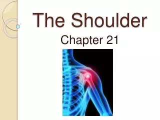
Chapter 22: The Shoulder Complex. Jennifer Doherty-Restrepo, MS, LAT, ATC Academic Program Director, Entry-Level ATEP Florida International University Acute Care and Injury Prevention. Introduction. The shoulder is an extremely complicated region of the body

E N D
Chapter 22: The Shoulder Complex Jennifer Doherty-Restrepo, MS, LAT, ATC Academic Program Director, Entry-Level ATEP Florida International University Acute Care and Injury Prevention
Introduction • The shoulder is an extremely complicated region of the body • Joint with a high degree of mobility, but, not without compromising stability • Involved in a variety of overhead activities relative to sport • Susceptible to a number of repetitive and overused type injuries
Functional Anatomy • Great mobility, limited stability • Round humeral head articulates with flat glenoid • Rotator cuff and long head of the biceps provide dynamic stability during overhead motion • Supraspinatus compresses the humeral head • Other rotator cuff muscles depress the humeral head Integration of the capsule and rotator cuff • Scapula stabilizing muscles also provide dynamic stability • Relationship with the other joints of the shoulder complex and the G-H joint is critical
Functional Anatomy • Scapulohumeral Rhythm • Movement of scapula relative to the humerus • Initial 30 degrees of G-H abduction • Does not incorporate scapular motion • Setting phase • 30 to 90 degrees of G-H abduction • Scapula abducts and upwardly rotates 1 degree for every 2 degrees of humeral elevation • Above 90 degrees of G-H abduction • Scapula and humerus move in 1:1 ratio
Prevention of Shoulder Injuries • Proper physical conditioning is key • Sport-specific conditioning • Strengthen through a full ROM • Warm-up should be used before explosive arm movements are attempted • Contact and collision sport athletes should receive proper instruction on falling • Protective equipment • Proper mechanics
Assessment of the Shoulder • History • What is the cause of pain? • Mechanism of injury? • Previous history? • Location, duration and intensity of pain? • Creptitus, numbness, distortion in temperature • Weakness or fatigue? • What provides relief?
Observation Elevation or depression of shoulder tips Position and shape of clavicle Position of head and arms Acromion process Biceps and deltoid symmetry Postural assessment (kyphosis, lordosis, shoulder) Scapular elevation and symmetry Scapular protraction or winging Muscle symmetry Scapulohumeral rhythm Assessment of the Shoulder
Spine of scapula Scapular vertebral border Scapular lateral border Scapular superior angle Scapular inferior angle Palpation: Bony Tissue • Sternoclavicular joint • Clavicular shaft • Acromioclavicular joint • Coracoid process • Acromion process • Humeral head • Greater and lesser tuberosity • Bicipital groove
Sternoclavicular, acromioclavicular, and coracoclavicular ligaments Rotator cuff muscles and tendons Subacromial bursa Sternocleidomastoid Biceps and tendon Coracoacromial ligament Glenohumeral joint capsule Deltoid Rhomboids Latissimus dorsi Serratus Anterior Levator scapulae Trapezius Supraspinatus Infraspinatus Teres major and minor Palpation: Soft Tissue
Special Tests • Active, Passive, and Resistive ROM • Flexion and extension • Abduction and adduction • Internal and external rotation • Manual Muscle Testing • Shoulder muscles and scapular stabilizers • RROM and Break tests
Special Tests: SC Joint Instability • Assesses sternoclavicular joint instability • Athlete in seated position • Apply pressure to the SC joint anteriorly, superiorly, and inferiorly • Determine stability or pain associated with a joint sprain
Special Tests: AC Joint Instability • Assesses acromioclavicular joint instability • Athlete in seated position • Palpate for displacement of acromion and distal head of clavicle • Apply pressure in all 4 directions • Determine stability or pain associated with a joint sprain
Special Tests: GH Joint Instability • Assesses glenohumeral joint instability • Special tests • Anterior and Posterior Drawer Tests • Sulcus Test • Clunk Test • Anterior and Posterior Apprehension Tests • Relocation Test
Anterior and Posterior Apprehension Tests • Anterior Apprehension Test • Posterior Apprehension Test
Relocation Test • Uses external rotation and posteriorly directed pressure to allow for increased external rotation
Special Tests: Impingement • O’Brien Test (Active Compression Test) • Flexion of GH joint to 90 degrees and horizontally adduction to 15 degrees • Passively place humerus into full IR and ER • If pain results with internal rotation but decreases with external rotation and if clicking is present, possible SLAP lesion • Pain in AC joint may indicate AC joint pathology
Special Tests: Impingement • Neer’s Test • Assesses impingement of soft tissue structures • Positive test is indicated by pain and grimace
Special Tests: Impingement • Hawkins-Kennedy Test • Assesses impingement of soft tissue structures • Positive test is indicated by pain and grimace
Special Tests: Rotator Cuff • Drop Arm Test • Assesses supraspinatus muscle weakness or tears • Athlete abducts shoulder and gradually lowers to starting position • Inability to lower arm slowly and controlled will indicate torn supraspinatus
Special Tests: Rotator Cuff • Empty Can Test • Place shoulder in position of 90 degrees of shoulder flexion, IR, and 30 degrees of horizontal abduction • Apply downward pressure • Assesses supraspinatus muscle weakness or tears
Special Tests: Serratus Anterior • Wall Push-up • Observe for winging scapula • Assesses for serratus anterior weakness • Could indicate injury to long thoracic nerve
Special Tests: Biceps • Yergason’s Test • Determines presence of biceps irritation and possible subluxation of biceps tendon • Speed’s Test • Determines presence of biceps irritation and possible subluxation of biceps tendon • Ludington’s Test • Assesses for possible rupture of biceps • Palpate alternating contractions of each biceps
Special Tests: Thoracic Outlet Syndrome • Adson’s Test • Assesses for anterior scalene syndrome • Compression of subclavian artery by scalenes • Athlete looks toward extended arm and takes a deep breath • Palpate radial pulse • Disappearance of pulse indicates a positive test
Special Tests: Thoracic Outlet Syndrome • Roo’s Test • Assesses for costoclavicular syndrome • Compression of subclavian artery between clavicle and first rib • Athlete assumes military brace position and turns head in opposite direction • Athlete opens and closes hand for 3 minutes Palpate radial pulse • Test is positive if… • Pulse disappears • Grip strength decreases
Special Tests: Thoracic Outlet Syndrome • Allen’s Test • Assesses for hyperabduction syndrome • Determines if pressure from pectoralis minor is compressing brachial plexus and subclavian artery
Specific Injuries • Clavicular Fractures • Etiology • MOI = fall on outstretched arm, fall on tip of shoulder, or direct impact • Occurs primarily in middle third • Signs and Symptoms • Athlete supports arm, head tilted towards injured side with chin turned away • Clavicle may appear lower • Palpation reveals pain, swelling, deformity, and point tenderness
Clavicular Fractures (continued) • Management • Closed reduction - sling and swathe immediately • Refer for X-ray • Immobilize with brace for 6-8 weeks • After removal of brace, rehabilitation includes: • Joint mobilizations • Isometric exercises • Use of a sling for 3-4 weeks • May require surgical treatment
Specific Injuries • Scapular Fractures • Etiology • MOI = direct impact or force transmitted up through humerus • Signs and Symptoms • Pain during shoulder movement • Swelling and point tenderness • Management • Sling immediately and refer for X-ray • Use sling for 3 weeks then begin PRE exercises
Specific Injuries • Fractures of the Humerus • Etiology • MOI = direct impact, force transmitted up through humerus, or fall on outstretched arm • Proximal fractures occur due to direct blow • Dislocations occur due to fall on outstretched arm • Epiphyseal fractures are more common in young athletes and occur due to direct blow or indirect blow traveling along long axis of humerus
Specific Injuries • Fractures of the Humerus (continued) • Signs and Symptoms • Pain, swelling, point tenderness, decreased ROM • Management • Immediate application of splint • Refer for X-ray • Treat for shock
Specific Injuries • Sternoclavicular Sprain • Etiology • MOI = indirect force or blunt trauma • Signs and Symptoms • Grade 1 - pain and slight disability • Grade 2 - pain, subluxation deformity, swelling, point tenderness, and decreased ROM • Grade 3 - gross deformity (dislocation), pain, swelling, and decreased ROM • Possibly life-threatening if dislocates posteriorly
Specific Injuries • Sternoclavicular Sprain (continued) • Management • RICE • Refer for reduction if necessary • Immobilize for 3-5 weeks • After immobilzation period, begin PRE exercises
Specific Injuries • Acromioclavicular Sprain • Etiology • MOI = direct blow (from any direction) or upward force from the humerus • Graded from 1 - 6 according to severity of injury • Signs and Symptoms • Grade 1 - point tenderness, pain with movement • No disruption of AC joint • Grade 2 - tear or rupture of AC ligament, pain, point tenderness, and decreased ROM (abd/add) • Partial displacement of lateral end of clavicle
Acromioclavicular Sprain (continued) • Signs and Symptoms • Grade 3 - rupture of AC and CC ligaments • AC joint separation • Grade 4 - posterior dislocation of clavicle • Grade 5 – rupture of AC and CC ligaments, tearing of deltoid and trapezius attachments, gross deformity, severe pain, decreased ROM • Grade 6 - displacement of clavicle behind the coracobrachialis
Acromioclavicular Sprain (continued) • Management • Ice, sling and swathe • Referral to physician • Grades 1 – 3: non-operative treatment • 1 - 2 weeks of immobilization • Grades 4 – 6: surgery required • Aggressive rehab is required for all AC sprains • Joint mobilizations, flexibility exercises, and PRE exercises should occur immediately • Progress as tolerated – no pain and no additional swelling • Padding and protection may be required until pain-free ROM returns
A: Grade 1 • B: Grade 2 • C: Grade 3 • D: Grade 4 • E: Grade 5 • F: Grade 6
Specific Injuries • Glenohumeral Joint Sprain • Etiology • MOI = forced abduction and/or external rotation; or a direct blow • Signs and Symptoms • Pain during movement • Especially when re-creating the MOI • Decreased ROM • Point tenderness
Specific Injuries • Glenohumeral Joint Sprain (continued) • Management • RICE for 24-48 hours • Sling • After hemorrhaging subsides, modalities may be utilized along with PROM and AROM exercises to regain full ROM • When full ROM achieved without pain, PRE exercises can be initiated • Must be aware of potential development of chronic conditions (instability)