Download
1 / 35
380 likes | 715 Views
Evaluation of Patients in Coma. Liam Durcan MD FRCPC Department of Neurology and Neurosurgery. What We’ll Cover. Basic definitions Key exam points Epidemiology of Coma Coma mimics. What we won’t talk about. Brain death/ chronic vegetative state toxidromes really complex neuroanatomy

E N D
Evaluation of Patients in Coma Liam Durcan MD FRCPC Department of Neurology and Neurosurgery
What We’ll Cover • Basic definitions • Key exam points • Epidemiology of Coma • Coma mimics
What we won’t talk about • Brain death/ chronic vegetative state • toxidromes • really complex neuroanatomy • Exhaustive lists of causes • Basic Resuscitative Care
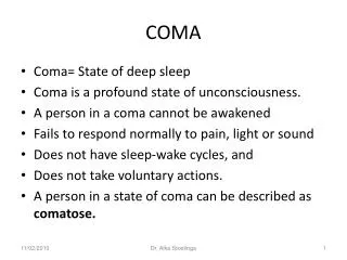
Definitions • Coma: “Unarousable unresponsiveness in which the subjects lie with eyes closed” • Plum and Posner- Diagnosis of Stupor and Coma • Other terms: obtundation, stupor • fallen out of favour because of imprecision • descriptive methods favoured
Consciousness • Two components of conscious behavior • content- the sum of cognitive and affective function • arousal- appearance of wakefulness • Content depends on arousal but normal arousal does not guarantee normal content
Really Simple Neuroanatomy • Arousal: where is it localized? • Ascending Reticular Activating System (ARAS) ‘core of the brainstem’ • receives input from numerous somatic afferents • projects to midline thalamic nuclei (which are in a circuit with cortical structures) and the limbic system
ARAS • ARAS acts as a gating system, increasing or decreasing thalamic inhibitory influence on the cortex • alters effect of sensory stimuli ascending • alters descending cortical stimulation
Demands of Arousal • Function of ARAS-Thalamic-Cortical system depends on: • anatomic integrity of structures • metabolic integrity (circulatory integrity) • communicative integrity (neurotransmitter function)
Coma Fact Number One • Coma implies dysfunction of: • ARAS or • Both hemi-cortices • Anatomically, this means • central brainstem structures (bilaterally) from caudal medulla to rostral midbrain • both hemispheres
Epidemiology of Coma • Plum and Posner 1982 • 500 consecutive cases of coma • 101 supratentorial (44/101 ICH) • 65 subtentorial lesions (40/65 brainstem infarcts) • 326 diffuse or metabolic brain dysfunction • 149 drug intoxication
Clues from History • Onset of symptoms • sudden onset • fluctuations • Associated neurologic symptoms • Medications
Neurologic Exam • Cornerstone of assessment • Descriptive, systematic • Reference point for serial assessment
Exam goals • Primary CNS event versus secondary • Implications: • short and long-term outcome • investigations
Breathing • Abnormalities of respiration can help localize but almost always in the context of other signs • Central-reflex Hyperpnea (midbrain-hypothalamus) • Apneustic, cluster, Ataxic (Lower pons) • Loss of automatic breathing (medulla)
Cranial Nerve Exam • Systematic assessment of brainstem function via reflexes • Cranial Nerve Exam • Pupillary light response (CN 2-3) • Occulocephalic/calorics (CN 3,4,6,8) • Corneal reflex (CN 5,7) • Gag refelx (CN 9,10)
.Pupillary Light Responses • Afferent Limb: Optic Nerve • Efferent Limb: Parasympathetics via occulomotor • Midbrain integrity/ tectum • Uncal Herniation (3rd nerve dysfunction) • Pupillary resistance to insult
Pupillary Light Responses • Be aware of drug effects • Systemic and Local • Avoid ‘PERLA’ • State size, before and after light stimulation • Specify right and left
Pupils: Localizing Value • Pons-pinpoint pupils • Symp. Dysfinction plus parasymp.irritation • Midbrain-Large fixed pupils unresponsive to light, hippus • Horner’s- symp.dysfunction • Unilateral dilation- parasymp. Dysfunction usually due to 3rd nerve lesion
Ciliospinal Reflex • 1-2 mm pupillary dilatation evoked by noxious cutaneous stimulation • More prominent in sleep or coma than during wakefulness • Test integrity of symp.pathways in comatose patients • Not particularly useful in evaluating brainstem function
Corneal Reflex • Afferent: Trigeminal Nerve • Efferent: Third Nerve (Bell’s Phenomenon and Facial Nerve (Eye closure) • Tests dorsal midbrain (Bell’s) and pontine integrity (Eye closure)
Eye Movements • Before maneuvers attempted note resting position • Midline • Deviation suggests frontal/pontine damage • Conjugate • Dysconjugance suggests CN abn. • Moving • Roving, dipping, bobbing
Occulocephalic/ Calorics • Same reflex elicited differently • Afferent: Eighth nerve • Efferent: 3,4,6 via MLF and PPRF • Occulocephalics may also involve proprioceptive afferents from the neck
Occulcephalic Reflex • Brisk rotation of head with eyes held open • Watch for contraversive movements • Next: • Flexion: eyes deviate up and eyelids open (doll’s head phenomenon) • Extension:eyes deviate downward
Caloric reflex • Ensure TM integrity • Elevation of head to 30 degrees (so that lateral semicircular canal is vertical) • Instillation of up to 120 ml of ice water • Awake: deviation toward,nystagmus away • Comatose: deviation toward • Wait 5 minutes, do other ear
Calorics • Watch for conjugance of deviation • To test vertical eye movements • Both ears, cold water-downward gaze • Both ears, warm water-upward gaze
Gag Reflex • Afferent: Glossopharyngeal • Efferent: Vagus • Taken in context of other findings
Motor Exam • Assess tone, presence of asterixis • Response to painful stimuli • none • abnormal flexor • abnormal extensor • normal localization/withdrawal • Avoid use of decerebrate/ decorticate
Reflexes • Brainstem • Deep tendon • Biceps, brachioradialis, triceps • Patellar, Achilles • Plantar Responses • Superficial skin • Abdominal, cresmasteric
Uncal herniaiton • Expanding lesions in lateral middle fossa • Compression of hippocampal gyrus over free edge of tentorium • Three stages described • Early third nerve • Late third nerve • Midbrain-Upper pons stage
Goals in Emergency • Primary Neurological Process? • evidence of raised ICP • focal findings, especially that implicate brainstem structures • Secondary Processes • signs of infection, toxic/metabolic processes • relative lack of focality
Coma Mimics • Akinetic mutism • ‘Locked-in’ syndrome • Catatonia • Conversion reactions
Akinetic Mutism • Silent, immobile but alert appearing • Usually due to lesion in bilateral mesial frontal lobes, bilateral thalamic lesions or lesions in peri-aqueductal grey (brainstem)
“Locked-In’ Syndrome • Infarction of basis pontis (all descending motor fibers to body and face) • May spare eye-movements • Often spares eye-opening • EEG is normal or shows alpha activity
Catatonia • Symptom complex associated with severe psychiatric disease with: • stupor, excitement, mutism, posturing • can also be seen in organic brain diease: encephalitis, toxic and drug-induced psychosis
Conversion reactions • Fairly rare • Occulocephalics may or may not be present • The presence of nystagmus with cold water calorics indicates the patient is physiologically awake • EEG used to confirm normal activity