Download
1 / 26
360 likes | 1.5k Views
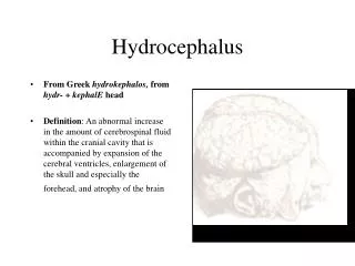
Emergency Evaluation of Hydrocephalus Shunt Patients. The Society of Neurological Surgeons Bootcamp. Communicating vs. Obstructive Hydrocephalus. Communicating Hydrocephalus All 4 ventricles are enlarged Causes: IVH of prematurity (grade III/IV), adult IVH, aneurysmal SAH, meningitis

E N D
Emergency Evaluation of Hydrocephalus Shunt Patients The Society of Neurological Surgeons Bootcamp
Communicating vs. ObstructiveHydrocephalus • Communicating Hydrocephalus • All 4 ventricles are enlarged • Causes: IVH of prematurity (grade III/IV), adult IVH, aneurysmal SAH, meningitis • May do lumbar puncture • Obstructive Hydrocephalus • Dilatation of lateral and third ventricles with small, compressed or normal size 4th ventricle • Asymmetry or enlargement of lateral ventricle when obstruction is at Foramen of Monro ( e.g. colloid cyst) • Posterior fossa mass lesions (tumor, ICH, cyst), intraventricular mass lesions (tumor, IVH, cyst), aqueductal stenosis • Do NOT do lumbar puncture
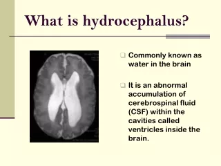
Communicating Hydrocephalus • Enlargement of lateral, 3rd, and 4th ventricles • Note sulcal effacement, temp horns, rounded 3rd, and enlarged 4th
Obstructive Hydrocephalus • Aqueductal stenosis • Note enlarged frontal horns, temporal tip dilation, rounded 3rd but small or normal 4th ventricle
Shunt Technology • Pressure differential valves • Antisiphon valves • Flow regulated valves • Programmable valves OSV
CSF Shunt Malfunction:Infants • Progressive macrocephaly • Tense anterior fontanelle • Sutural splaying • Downgaze, lid retraction • Esotropia (VIth nerve palsy)
CSF Shunt Malfunction:Children • Developmental delay • Decline in school performance (esp. verbal IQ) • Visual loss
Radiology • Compare ventricular size to “well”baseline • Infants: Trans-fontanelle ultrasound • CT • MRI • Shunt x-ray series • Disconnection or fracture of tubing
Invasive Studies • CSF shunt tap • Assess flow and pressure (although proximal obstruction may commonly interfere with accuracy) • Send CSF for GS/Cx, Glu/Pro, cell counts if infection suspected • Relieve pressure if obstructed distally • Radionuclide shuntogram • Assess proximal and distal flow • Ventricular reflux and outflow each correlate with appropriate function (but test is imperfect) • Intracranial pressure monitoring
CSF Shunt Infection • Therapy • Externalize shunt • Change hardware • Antibiotics • Consider LP • Organisms • Staph. Epi (40%) • Staph. Aureus (20%) • Gram Negatives • Diptheroids • Yeast
Differential Diagnosis of Shunt Infection • Gastroenteritis • Often associated with sick contacts, diarrhea • Otitis • May often be detected on physical examination • Urinary tract infection • Important to differentiate from colonization in spina bifida patients
CSF Shunt Complications:Mechanical Failure • Blockage • Choroid plexus • Ependyma • Fracture • Disconnection • Valve failure
CSF Shunt Complications:Mechanical Failure • Distal failure • Kinked tubing • Malabsorption • Pleural effusion • Cor pulmonale • Shunt nephritis
CSF Shunt Complications:Abdominal failure • Umbilical hernia • Extra-peritoneal catheter • Bowel perforation
CSF Shunt Complications:Overdrainage • Postural (Low pressure) headache • Subdural hygroma • Craniostenosis
CSF Shunt Complications:Hemorrhage • Parenchymal damage • Raised ICP • IVH: Valve obstruction • Ependymal adhesions and multicompartmental hydrocephalus
Shunt Evaluation Protocol:History • History • Hydrocephalus etiology • Exact date of last tap or revision • Symptoms of last failure • Seizure disorder? • Latex allergy? • Current Symptoms • Headache • Severity/location • Positional • Morning • Mental status changes • Fever • Shuntalgia • Nausea/vomiting • Intercurrent illness
Shunt Evaluation Protocol:Diagnostic Studies • Non-contrast head CT scan (shunt protocol) or‘quick brain’ MRI • Shunt x-ray series • Abdominal ultrasound, if indicated • Shunt tap, if indicated • Formal skin preparation • 25g butterfly needle: test OP and valsalva (OP may be obscured by proximal obstruction) • CSF sample for GS/Cx, Cell count, Glu/Prot
Shunt Evaluation Protocol:Admission • Immediate intervention for: • Definite, acute malfunction • Pain • Infection • Bradycardia • Decreased mental and/or vision • Cardiorespiratory monitoring • Frequent neurological checks • NPO except meds • Anti-microbial shampoo • Consider steroid prep for latex allergy
Conclusions • Involve experienced team members in significant care decisions • When in doubt, keep the patient for observation • Listen to parents • Myelomeningocele patients may have protean forms of presentation and increased risk for sudden deterioration • Remember that, above all, shunt malfunction is a clinical diagnosis, supported by imaging studies and other data
Case 1 • History • 6 y.o. with post-hemorrhagic hydrocephalus • 3 days progressive fever and malaise • Intermittent right sided headaches • Last revision 3 years ago for obtundation
Case 1 • Physical Examination • Irritable • Neurological exam non-focal • Temperature 102.5 F. • Inflamed right tympanic membrane with effusion • Imaging • Axial imaging: ventricles unchanged from last well scan • Shunt x-rays without disconnection • Diagnosis • Otitis media • No surgical intervention
Case 2 • History • 10 y.o. with myelomeningocele and hydrocephalus • One week of progressive frontal headaches and neck pain • One day of vomiting • Mother states these are typical malfunction symptoms • Last revision distant • Physical Examination • Alert • Baseline • No papilledema • Radiology • Axial imaging unchanged from well baseline (small ventricles) • Shunt x-rays without disconnection
Case 2 • Diagnosis • VP shunt malfunction • Total proximal shunt obstruction was observed at surgery
Case 3 • History • 10 y.o. brought to E.R. by ambulance, obtunded • EMT: “Has a shunt for hydrocephalus; had headaches at home for last few days” • Physical Examination • Unresponsive • RR 15, labored • HR 70 • Pupils 4 mm, sluggish • Frontal valve-reservoir palpable
Case 3 • Diagnosis • Severe ventricular shunt malfunction • Treatment • Neurosurgeon attempts to drain CSF; shunt tap is dry • 1 gram/kg mannitol is given • E.R. Course • Intubated • During CT, heart rate drops to 40