Download
1 / 46
490 likes | 661 Views
Transport in Humans (and other Mammals). The Circulatory System. Cardiovascular System. Systemic circulation. Pulmonary circulation. HEART. LUNGS. BODY. Double Circulatory System. Consists of Heart, Lungs and Vessels. The Heart. Weighs approx 300g

E N D
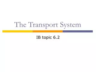
Transport in Humans (and other Mammals) The Circulatory System
Cardiovascular System Systemic circulation Pulmonary circulation HEART LUNGS BODY Double Circulatory System • Consists of Heart, Lungs and Vessels
The Heart • Weighs approx 300g • Thick muscular layer (myocardium) made of Cardiac muscle • Has it’s own blood supply • covered in capillaries which get blood from coronary arteries • Myogenic- self exciting • Continuous rhythm (can be altered)
Two upper thin walled chambers – atria • Two lower thick walled chambers – ventricles • Atria collect blood from the body – attached to veins • Ventricles send blood to the body – attached to arteries
The Heart • Right side – deoxygenated • Left side – oxygenated • Separated by a thick walled septum • Right atrium and ventricle connected by tricuspid valve • Left atrium and ventricle connected by bicuspid (mitral) valve • Cuspid valves held shut by Chordae Tendinae • Left ventricle is thicker than the right
Vessels of the Heart • Pulmonary artery* • Pulmonary vein • Superior and Inferior Vena cava • Aorta* • *Semi-lunar valves
Diagram of the Heart pg 13
Blood Flow Through the Heart • Vena Cava – Superior/Inferior • Right Atrium • Tricuspid Valve • Right ventricle • Pulmonary Artery LUNGS
Blood Flow Through the Heart • Pulmonary veins • Left Atrium • Bicuspid/Mitral/Atrioventricular valve • Left Ventricle • Aorta MAJOR ORGANS
Control of the Heart • Myogenic-Initiated from inside the heart as opposed to nervous stimulus outside • Initial Stimulus originates in the Sinoatrial node (SA node) • A pacemaker that determines heart rate • Wave of excitation across both atria causes them to contract
Control of the Heart • Atrioventricular node (AV node) • Sends waves of excitation along Purkinje Fibres which collectively make up the Bundle of His. Along septum, radiate upwards • Causes ventricles to contract
Cardiac cycle • Systole -contraction of heart • Diastole -Relaxation/filling of the heart • Both atria contract at the same time • Called …………………. • Both ventricles contract at same time • Called …………………. • Relaxation of atria and ventricles • Called ……………………….
Cardiac cycle • Remind me…. What is it?
Vessels of the Cardiovascular System Arteriole Artery Capillary Vein Venuole
Vessels of the Cardiovascular System • Arteries – Arterioles – Capillaries - Venuoles - Veins • Different structures and functions
Arteries • Carry blood away from the heart • Blood in arteries is under high pressure • Their structure is related to their function • Round in structure with relatively thick walls composed of three layers
Arteries • Tunica Intima – Single layer of endothelial cells • Tunica Media – A thick layer containing elastic fibres and muscle tissues. Closer to heart more elastic fibres those further away have more muscle fibre • Tunica Externa – Contains collagen fibres for strength
Blood vessels – the arteries TUNICA MEDIA TUNICA EXTERNA TUNICA INTIMA ( LUMEN
Veins • Carry blood back to the heart (generally deoxygenated) • Blood now flowing slowly, smoothly and under low pressure (structure related to this function) • Walls are relatively thin compared with arteries • Still same three layers but the tunica media differs in size – relatively narrow with few muscle fibres and elastic fibres • Semi Lunar valves present along length
Blood vessels – the veins TUNICA MEDIA TUNICA INTIMA TUNICA EXTERNA LUMEN
Capillaries • Link the arterial and venous blood supplies • Blood flow is slow, blood pressure is falling and is non-pulsatile • Capillaries form vast networks in all tissues and organs • Composed of only the Tunica Intima, only a single layer of endothelial cells, no elastic fibres and no muscle tissue
Capillaries • Exchange of materials between the blood and the body takes place in the capillary bed
Blood vessels – the capillaries LUMEN ONE CELL THICK
Time for an exam question page 13
Components of Blood • Blood is made up of: • Variety of cells • Suspended in fluid – Plasma • All blood cells develop from stem cells in bone marrow
Plasma Transports everything. Blood cells. Digested foods (glucose). Waste. Hormones 90% water, 10% solutes 3 Proteins: Globulins, Albumins and Fibrinogen immune clotting
Red Blood cells Erythrocytes Red Blood cells carry oxygen around the body in haemoglobin. They have no nucleus thus leaving more space for oxygen
White blood cells Leucocytes Fight against disease. Destroys bacteria by using antibodies. Fights toxins by using antitoxins. Kills foreign microbes by consuming them.
White blood cells cont. • WBC’s divided into 2 groups: • GRANULOCYTES • AGRANULOCYTES • Granulocytes have granules in cytoplasm & multilobed nucleus. 3 types: • Neutrophils • Eosinophils • Basophils • Agranuloycytes are NOT granular or lobed • Monocytes • lymphocytes
Platelets Thrombocytes Platelets are tiny fragments in the body that help blood clot at wounds.
Transport of gases • Oxygen combines with Haemoglobin: Haemoglobin + oxygen oxyhaemoglobin • Reversible so O2 is available to body tissues. • Each g of haemoglobin can combine with 1.34cm3 of oxygen. • See WJEC (oxygen dissociation)
Oxygen Dissociation Curves • Concentration of gases are measured in partial pressures • As haemoglobin is exposed to increasing ppO2, haemoglobin takes it up until saturated • Each haemoglobin can take up ___ molecules O2
Curve shape depends on number of factors: pp CO2 Temp pH Increasing ppCO2 shifts curve right, Bohr Effect due to decrease in affinity of haemoglobin to O2 Means oxyhaemoglobin releases more O2 as CO2 rises ie when cells are respiring more Oxygen Dissociation Curves
Fetal Haemoglobin • Haemoglobin F has a higher affinity for oxygen than adult haemoglobin • Means it is able to gain oxygen from mothers haemoglobin
Myoglobin • Myoglobin is a pigment found in muscle and hearts of mammals • Combines reversibly with Oxygen • Takes up oxygen more readily than haemoglobin • Blood reaching muscle transfers O2 to myoglobin acting as a temp store • Myoglobin gives up its O2 when ppO2 drops to very low value, eg after exercise
CO2 • See WJEC (transport of gases) • CO2 produced in respiration diffuses into blood and into RBCs • In RBCs the CO2 combines with H2O to form carbonic acid (H2CO3) – enzyme carbonic anhydrase • Carbonic acid then dissociates to hydrogencarbonate ions CO2 + H2O H2CO3 H+ + HCO3-
CO2 • Hydrogencarbonate ions diffuse into plasma in exchange for Cl- ions • CO2 reacts with haemoglobin and other proteins to form carbamino compounds • CO2 can be transported in blood 3 ways: • Hydrogencarbonate ions • Carbamino compounds • Simple solution, dissolved in plasma
Tissue Fluid • Fluid that leaks out of capillaries is called tissue fluid • It fills spaces between body cells • It contains: • Water Glucose • Amino acids Fatty acids • Glycerol mineral ions • vitamins • Dissolved gases
Blood pressure at arteriole end of capillaries (5.3kPa) forces water out • This hydrostatic pressure must be greater than the osmotic pressure drawing water in. • Plasma Proteins stay in capillaries (so low ψ) and the hydrostatic pressure keeps dropping as the blood passes through the capillary beds.
At venular end, pressure has dropped to less than that of the osmotic effect of the proteins (1.3kPa) • Water drawn back in to capillaries (99% of it anyway) • Excess fluid and some waste products like urea enter lymph system and are returned via that pathway.