Download
1 / 88
950 likes | 1.54k Views
Functional Human Physiology for Exercise and Sport Sciences The Urinary System: Renal Function. Jennifer L. Doherty, MS, ATC Department of Health, Physical Education, and Recreation Florida International University. Functions of the Urinary System.

E N D
Functional Human Physiologyfor Exercise and Sport Sciences The Urinary System: Renal Function Jennifer L. Doherty, MS, ATC Department of Health, Physical Education, and Recreation Florida International University
Functions of the Urinary System • The kidneys remove metabolic wastes from the blood and excrete them to the outside of the body in the form of urine • The careful regulation of renal activity keeps blood composition and body fluids within normal limits
The kidneys also… • Maintain electrolyte and acid-base balance in body fluids • Regulate plasma pH by regulating the concentration of bicarbonate ions and hydrogen ions • Regulate the volume, composition, and pH of blood • Regulate plasma osmolarity and chemical composition • Assist in the regulation of BP • Regulate plasma volume and produces renin to regulate BP • Assist in the regulation of RBC production • Regulate RBC production by producing erythropoietin to stimulate RBC formation in bone marrow • Assist in the regulation of Ca++ absorption • Metabolize vitamin D to its active form, which affects the rate of Ca++ absorption from the small intestines
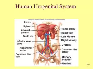
Anatomy of the Urinary System Structures of the urinary system • Kidneys (2) • Form urine • Renal arteries and veins • Ureters (2) • Tubes for transport of urine from the kidneys to the bladder • Urinary bladder (1) • Storage reservoir for urine • Urethra (1) • Transport tube for urine to the outside of the body
Microscopic Anatomy of the Kidneys • The nephron is the structural and functional unit of the kidney. • Each kidney contains about one million nephrons • Nephrons consist of: • The renal corpuscle • Proximal convoluted tubule • Loop of Henle • Distal convoluted tubule • Collecting duct • Empties urine into the minor calyx
The Nephron: Renal Corpuscle • The renal corpuscle consists of… • Glomerulus • Glomerular Capsule
The Nephron: Renal Corpuscle • The Glomerulus is the filtering unit of the nephron • Tangled cluster of capillary beds lying between the afferent and efferent arterioles • Contained within the glomerular capsule
The Nephron: Renal Corpuscle • The Glomerular Capsule (Bowman’s capsule) is a thin walled, cup-shaped structure surrounding the glomerulus • It leads to the renal tubule • It receives fluid that filters through the glomerulus
Basic Renal Exchange Processes • Nephrons function to… • Remove wastes from the blood • Regulate water and electrolyte concentrations • Urine is the end product of these functions • The following 3 exchange processes occur within the nephrons • Glomerular Filtration • Reabsorption • Secretion
Glomerular Filtration • This is the beginning of urine formation • Glomerular capillaries are extremely permeable compared to systemic capillaries • Hydrostatic and Osmotic Pressure Gradients • Greater inside glomerular capillaries • Forces water and dissolved solutes to leave the blood plasma in the glomerular capillaries and cross the glomerular membrane into the glomerular (Bowman’s) capsule • Filtrate • Water and dissolved solutes in the glomerulus
Glomerular Filtration The glomerular membrane • Separates glomerular capillary blood from the glomerular capsule space • Contains many small pores that allow almost all materials to pass through the membrane • Exceptions: formed elements
Glomerular Filtration Glomerular filtrate • Contains water and dissolved solutesthat have been filtered from the blood plasma in the glomerular capillaries and collected by the glomerular capsule • Similar to tissue fluid containing water, glucose, amino acids, urea, uric acid, creatine, creatinine, sodium, chloride, potassium, calcium, bicarbonate, phosphate, and sulfate ions • Will be processed by the renal tubules to form urine
Glomerular Filtration • A nonselective, passive process • Water and dissolved solutes from the blood plasma in glomerular capillaries are forced through the glomerular membrane by hydrostatic and osmotic pressure gradients • Water and dissolved solutes travel down their pressure gradients
Glomerular Filtration Glomerular Filtration Pressure (Net Filtration Pressure) • The net force acting to move materials out of the glomerulus (glomerular capillaries) and into the glomerular capsule • Filtration pressure is much higher in the glomerular capillaries compared to systemic capillaries because of: • The high permeability of the glomerular membrane • It is more permeable than systemic capillary membranes • High glomerular blood pressure (60 mmHg) • It is higher compared to systemic capillary blood pressure (41 mmHg)
Glomerular Filtration Starling Forces • Represent the overall effect of all the forces operating at the glomerular membrane
Glomerular Filtration: Starling Forces • Forces favoring filtration are the forces driving fluid and solutes out of the glomerular capillaries • Glomerular capillary hydrostatic pressure • PGC = 60 mmHg • The primary force pushing water and solutes out of the glomerular capillaries • Osmotic pressure in the glomerular (Bowman’s) capsule • πBC = 0 mmHg • Negligible since few plasma proteins are normally present in the glomerular capsule
Glomerular Filtration: Starling Forces • Forces opposing filtration are the forces driving fluid and solutes back into the glomerular capillaries • Glomerular (Bowman’s) capsule hydrostatic pressure • PBC = 15 mmHg • Exerted by the fluids within the glomerular capsule • Osmotic pressure in the glomerular capillaries • πGC = 29 mmHg • Due to the plasma proteins in glomerular blood
Glomerular Filtration Pressure • Glomerular Filtration Pressure Equation • Filtration pressure = (forces favoring filtration) - (forces opposing filtration) • Forces favoring filtration • (glomerular capillary hydrostatic pressure + capsular osmotic pressure) • Forces opposing filtratrion • (capsular hydrostatic pressure + glomerular capillary osmotic pressure) • Values • (60 mmHg + 0 mmHg) - (15 mmHg + 29 mmHg) = 16 mmHg
Glomerular Filtration Rate (GFR) • GFR = the amount of filtrate produced in the kidneys per minute • Normal values: • 125 ml/min (180 L/day) • GFR varies with the filtration pressure • All the factors that affect glomerular filtration pressure will affect the GFR • Glomerular capillary osmotic pressure • Glomerular capillary hydrostatic pressure • Glomerular capsule osmotic pressure • Glomerular capsule hydrostatic pressure
Glomerular Filtration Rate (GFR) • Glomerular capillary hydrostatic pressure • ↑ glomerular capillary hydrostatic pressure = ↑ GFR • Glomerular capsule osmotic pressure • ↑ glomerular capsule osmotic pressure = ↑ GFR • Glomerular capillary osmotic pressure • ↑ glomerular capillary osmotic pressure = ↓ GFR • Glomerular capsule hydrostatic pressure • ↑ glomerular capsule hydrostatic pressure = ↓ GFR
Glomerular Filtration Rate (GFR) • GFR varies with the rate of blood flow through the glomerular capillaries • To maintain a high GFR, blood must flow quickly through glomerular capillaries • Vasocontriction or vasodilation in the glomerular arterioles elicit changes in the glomerular filtration pressure • Changes in the glomerular filtration pressure effect GFR
Glomerular Filtration Rate (GFR) • Vasoconstriction of the afferent arterioles or vasodilation of the efferent arterioles • ↓ glomerular capillary hydrostatic pressure • ↓ GFR • Vasodilation of the afferent arterioles or vasoconstriction of the efferent arterioles • ↑ glomerular capillary hydrostatic pressure • ↑ GFR
Regulation of GFR • GFR remains relatively constant • May be ↑ or ↓ according to the body’s need • Mechanisms of regulation: • Intrinsic control (Autoregulation) • Myogenic regulation • Tubuloglomerular feedback • Extrinsic control • Renal blood flow • Exercise
Regulation of GFR: Intrinsic Control • The ability of the kidney to maintain a constant blood flow when arterial BP is changing • The ability of the kidneys to maintain a relatively constant GFR when mean arterial pressure is changing • This mechanism is effective over the "normal" range of arterial BP • 80 - 120 mmHg
Regulation of GFR: Intrinsic Control Myogenic Regulation • Related to the inherent property of smooth muscle to contract when stretched • ↑ mean arterial pressure = ↑ stretch of smooth muscle in the afferent arteriole walls stimulating vasoconstriction • Vasoconstriction of the arterioles causes a decrease in glomerular capillary hydrostatic pressure • This protects the delicate glomerular capillaries from high mean arterial pressures • Myogenic regulation is especially effective in the afferent arteriole
Regulation of GFR: Intrinsic Control Tubuloglomerular Feedback • Negative feedback system • GFR is regulated by changes in flow of tubular fluid past the macula densa • Specialized cluster of epithelial cells in the distal convoluted tubule near the afferent and efferent arterioles • Changes in Na+ andCl- concentration in the filtrate are detected by osmoreceptors in the macula densa
Tubuloglomerular Feedback Macula Densa Cells • Respond to changes in the Na+Cl- concentration in the filtrate in the distal convoluted tubule • ↓ Na+Cl- concentration = afferent arteriole vasodilation • Vasodilation of the afferent arteriole results in: • ↑ blood flow to the glomerular capillaries • ↑ glomerular filtration pressure • ↑ GFR • The opposite is also true
Tubuloglomerular Feedback Juxtaglomerular Cells • Contain mechanoreceptors that stimulate the juxtaglomerular cells to release renin in response to changes in mean arterial pressure • Renin is an enzyme that catalyzes a cascade of reactions in the bloodstream • Renin converts angiotensinogen → angiotensin I • Angiotensin converting enzyme (ACE) converts angiotensin I → angiotensin II • Angiotensin II is the most powerful vasoconstrictor in the body • Increases mean arterial blood pressure
Tubuloglomerular Feedback • Activation of the juxtaglomerular cells to release renin occurs when there is a decrease in mean arterial pressure • Usually when mean arterial pressure is less than 80 mmHg • Direct activation of juxtaglomerular cells • Achieved via the mechanoreceptors sending impulse through the sympathetic nervous system • Indirect activation of juxtaglomerular cells • Achieved via the macula densa cells which detect changes in Na+Cl- concentrations in the filtrate • Macula densa cells cause vasoconstriction or vasodilation, which alters mean arterial pressure as detected by the juxtaglomerular cells
Regulation of GFR: Extrinsic Control Renal Blood Flow • The sympathetic nervous system is able to override autoregulation of the kidneys • ↑ sympathetic input = ↓ GFR • Sympathetic input causes vasoconstriction of both afferent and efferent arterioles, thereby decreasing GFR
Regulation of GFR: Extrinsic Control Exercise • Exercise results in increased sympathetic nerve impulses • Sympathetic nerve impulses stimulate the adrenal medulla to release epinephrine, which stimulates… • Release of renin = ↑ mean arterial pressure • Vasoconstriction of the afferent arteriole = ↓ GFR
Reabsorption • The process of reclaiming fluid and solutes from the filtrate in the renal tubules • Reabsorption occurs in the peritubular capillaries • Solutes and water move from the lumen of the renal tubules back into the plasma • If reabsorption did not occur, a person would lose 1L of fluid in the urine in 8 min
Solute Reabsorption • Substances are selectively reabsorbed from the filtrate • Peritubular capillaries are specially adapted for the process of reabsorption • Under very low BP • Walls are very permeable • Reabsorption occurs throughout the renal tubule; however, most reabsorption occurs in the proximal convoluted tubule
Solute Reabsorption: Proximal Convoluted Tubule • Most reabsorption occurs in the proximal convoluted tubule • Proximal convoluted tubule contains epithelial cells with microvilli • Microvilli increase the surface area within the renal tubules
Solute Reabsorption: Proximal Convoluted Tubule • Solutes are moved from the tubule lumen, across the apical membrane, into the epithelial cells lining the tubule walls
Solute Reabsorption: Proximal Convoluted Tubule • Solutes then move out of the epithelial cells lining the tubule walls, across the basolateral membrane, into the peritubular space
Solute Reabsorption: Proximal Convoluted Tubule • From the peritubular space, solutes easily diffuse into the peritubular capillaries
Regional Specialization of the Renal Tubules: Proximal Tubule • Na+ ions are actively reabsorbed by active transport • Requires ATP • Reabsorption of Na+ establishes an electrical gradient for reabsorption of negatively charged ions • As positively charged Na+ ions are transported out of the filtrate, negatively charged ions accompany them via passive diffusion • Cl- • Bicarbonate (HCO3-) • Reabsorption of Na+ also establishes an osmotic gradient for the reabsorption of water • Water is passively reabsorbed by osmosis and returned to the systemic circulation by the peritubular capillaries
Regional Specialization of the Renal Tubules : Proximal Tubule • The mechanisms of reabsorption in the proximal tubule are so efficient that 70% of water and Na+ filtered is reabsorbed before the tubular fluid reaches the loop of Henle • Water and Na+ reabsorption are regulated by several hormones • At the end of the proximal convoluted tubule, the filtrate and the blood in the peritubular capillaries are isotonic (electrically neutral)