Download
1 / 33
380 likes | 571 Views
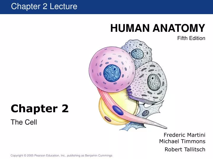
Chapter 2 Lecture. Chapter 2. The Cell. Frederic Martini Michael Timmons Robert Tallitsch. Introduction. Cell theory: Cells are the smallest structural units that perform all vital functions. The Study of Cells. The study of cells is cytology : Light microscopy

E N D
Chapter 2 Lecture Chapter 2 The Cell Frederic Martini Michael Timmons Robert Tallitsch
Introduction • Cell theory: • Cells are the smallest structural units that perform all vital functions.
The Study of Cells • The study of cells is cytology: • Light microscopy • Transmission electron microscopy • Scanning electron microscopy Figure 2.1a,b,c Different Technique, Different Perspective
Cellular Anatomy Cells have four types of component: Membranes Organelles Cytoplasm inclusions Figure 2.3 Anatomy of a Typical Cell
Cellular Anatomy Figure 2.4 A Flow Chart for the Study of Cell Structure
Cell Structure This movie reviews cell structure. Cell Structure PLAY
The Cell Membrane Figure 2.5 The Cell Membrane
The Cell Membrane • Major functions of the cell membrane can be described: • Regulation of exchange with the environment • Receptors and ID
Membrane Permeability: Passive Processes • Diffusion • Osmosis • Facilitated Diffusion Figure 2.6 Diffusion across the Cell Membrane
Membrane Permeability: Active Processes • Active transport uses enzymes and carrier proteins: • Ions pumps arecarrier proteins for charged particles. • Ions moved regularly by active transport include Na+, Ca2+,Mg2+,K+ • Ion pumps are specific. • An ion pump that moves two ions simultaneously in opposite directions is called an exchange pump.
Membrane Permeability: Active Processes Figure 2.7a Pinocytosis
Membrane Permeability: Active Processes Figure 2.7b Phagocytosis
Membrane Permeability: Active Processes Figure 2.8 Receptor-Mediated Endocytosis
Cytoplasm • The cytoplasm is the general term for the material inside the cell. • It is a fluid very high in protein. • The cytosol is the intracellular fluid: • It is high in potassium ions. • It contains an overall negative charge. • Transmembrane potential • It contains high concentrations of proteins. • Organelles are structures within the cyto-plasm that have a particular function and very distinct structure.
Nonmembranous Organelles • The cytoskeleton: • Microfilaments • Microtubules • Microvilli
Nonmembranous Organelles Figure 2.9 The Cytoskeleton
Nonmembranous Organelles • Centrioles • Cilia • Flagella Figure 2.10 Centrioles and Cilia
Nonmembranous Organelles • Ribosomes: • 60% RNA and 40% protein • Free ribosomes: • Float in the cytoplasm • Fixed ribosomes: • Are attached to the endoplasmic reticulum Figure 2.11 Ribosomes
Membranous Organelles • Mitochondria are double membraned organelles. • Cristae are the folds of the inner membrane • Inner fluid is the matrix. Figure 2.12 Mitochondria
Membranous Organelles • The nucleus is the control center of the cell. • Nucleoplasm • Nuclear envelope • Perinuclear space • Nuclear pores • Nuclear matrix
Membranous Organelles Figure 2.13a The Nucleus
Membranous Organelles: Nucleus • Chromosomes: • DNA wrapped around proteins called histones. • Nucleosomes • Chromatin Figure 2.14 Chromosome Structure
Membranous Organelles • The ER has four major functions: • Synthesis of all classes of macromolecules • Storage of the manufactured molecules • Transport of substances from on area of the cell to another • Enzymes in the lumen of the ER provide detoxification
Membranous Organelles Figure 2.15 The Endoplasmic Reticulum
Membranous Organelles • The three main functions of the Golgi apparatus: • Synthesis and packaging of secretions. • Packaging of enzymes for use in the cytosol. • Renewal and modification of the cell membrane. Figure 2.16b The Golgi Apparatus
Membranous Organelles • Lysosome function in three manners: • Fuse with phagosomes to digest solid materials. • Fuse with and recycle damaged organelles. • Sometimes rupture a process resulting in autolysis. Figure 2.18 Lysosomal Functions
Intercellular Attachment Figure 2.19 Cell Attachments
The Cell Life Cycle Figure 2.20 The Cell Life Cycle
DNA Replication Figure 2.21 DNA Replication
Interphase Figure 2.22a
Prophase Figure 2.22b,c
Metaphase and Anaphase Figure 2.22d,e
Telophase and Cytokinesis Figure 2.22f,g