Download
1 / 69
700 likes | 972 Views
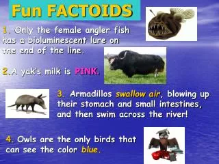
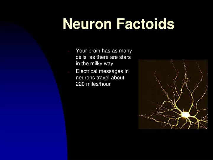
Neuron Factoids. Your brain has as many cells as there are stars in the milky way Electrical messages in neurons travel about 220 miles/hour. Anatomy and Physiology of the Neuron. I. Neuron Overview II. Membrane Characteristics A. Associated Proteins III. Membrane Potential A. Forces

E N D
Neuron Factoids • Your brain has as many cells as there are stars in the milky way • Electrical messages in neurons travel about 220 miles/hour
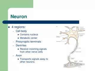
Anatomy and Physiologyof the Neuron I. Neuron Overview II. Membrane Characteristics A. Associated Proteins III. Membrane Potential A. Forces B. Equilibrium Potential C. Nernst Equation D. Action Potential
Introduction One can study the function of the nervous system at a number of levels of organization. In order to understand any level, one must understand the smallest functioning unit--the neuron.
Neuronal Communication • Even a simple behavior, like a reflex, requires collection, distribution, and integration of information. • The axonal membrane has properties that enable it to conduct a special type of signal, an action potential, leading to transference of communication.
Neuron Structure and Function • Inter- and intracellular communication
*Glia and NeuronsNeuroglia (“glue”): provide physical support Control nutrient flowCommunication with neurons PhagocytosisNeurons: Process informationSense environmental changes Communicate changes to other neuronsCommand body response
Astrocytes • Most numerous glia in the brain • Fill spaces between neurons • Influence neurite growth • Regulate ion concentration • Regulate glutamate • Communicate with neurons
Glia • Myelinating Glia • Oligodendroglia (in CNS) and Schwann cells (in PNS) • Insulate axons • Speed conduction
Glia • Myelinating Glia (Cont’d) • Node of Ranvier • Region where the axonal membrane is exposed • Other Non-Neuronal Cells • Microglia as phagocytes (immune)
The Neuron Doctrine • Histology • Study of tissue structure • The Nissl Stain • Facilitates the study of cytoarchitecture in the CNS
The Neuron Doctrine • Cajal’s Contribution • Neural circuitry • Neurons communicate by contact, not continuity • Neuron doctrine • Neurons adhere to cell theory
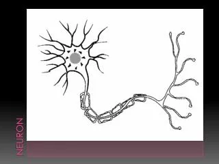
The Neuron Doctrine • Golgi-stain shows two parts of neurons: • Soma and perikaryon • Neurites: Axons and dendrites
Classifying Neurons • Classification Based on the Number of Neurites • Single neurite • Unipolar • Two or more neurites • Bipolar- two • Multipolar- more than two
Classifying Neurons • Classification Based on Dendritic and Somatic Morphologies • Stellate cells (star-shaped) and pyramidal cells (pyramid-shaped) • Spiny or aspinous
Classifying Neurons • Further Classification • By connections within the CNS • Primary sensory neurons, motor neurons, interneurons • Based on axonal length • Golgi Type I • Golgi Type II • Based on neurotransmitter type • e.g., – Cholinergic = Acetycholine at synapses
Neuron Classification Schemes • Neurons can be classified according to • Dendritic and Axonal Morphology: • Unipolar: one stalk that splits into two branches • Bipolar: one axon, one dendritic tree • Multipolar: one axon, many dendritic branches • Stellate and pyramidal cells • Spiny or aspinous • Golgi Type I and II • Function • Sensory neurons carry messages toward brain • Motor neurons carry messages to muscles • Interneurons connect cells • Neurotransmitter (NT) used by neuron • Effects of NT (excitatory vs. inhibitory)
The Prototypical Neuron • The membrane of the dendritic tree has specialized proteins called receptors in it to detect neurotransmitters released by other neurons into the synapse. • The cell body is the brain of the cell. • The axon is distinct in that it is critical for conduction of the nerve impulse. Key structures are the axon hillock and the terminal endings The axon terminal is different from the rest of the axon in 3 respects: 1. No microtubules extend into the terminal. 2. The terminal contains numerous small bubbles of membrane called synaptic vesicles, which are the storage location for neurotransmitters. 3. The terminal has numerous mitochondria, indicating that it requires a large amount of energy.
The soma • Nucleus • Gene expression • Rough Endoplasmic reticulum (ER) • Major site for protein synthesis • Ribosomes • Smooth ER and Golgi Apparatus • Sites for preparing/sorting proteins for delivery to different cell regions (trafficking) and regulating substances • Mitochondria • Site of cellular respiration (inhale and exhale) • Krebs cycle • ATP- cell’s energy source • Lysosomes
The Prototypical Neuron • The Axon • Axon hillock (beginning) • Axon proper (middle) • Axon terminal (end) • Differences between axon and soma • ER does not extend into axon • Protein composition: Unique
The Prototypical Neuron • The Axon • The Axon Terminal • Differences between the cytoplasm of axon terminal and axon • No microtubules in terminal • Presence of synaptic vesicles • Abundance of membrane proteins • Large number of mitochondria
The Prototypical Neuron • The Cytoskeleton • Not static • Internal scaffolding of neuronal membrane • Three “bones” • Microtubules • Microfilaments • Neurofilaments
Intracellular Proteins: Cytoskeleton • Microfiliments (proteins actin and mysin-growth cones) • Neurofiliments (Alzheimer’s Disease) • Microtubules (cell movement)
The Prototypical Neuron • The Axon • Axoplasmic transport • Anterograde (soma to terminal) vs. Retrograde (terminal to soma) transport
The Prototypical Neuron • Dendrites • “Antennae” of neurons • Dendritic tree • Synapse - receptors • Dendritic spines • Postsynaptic (receives signals from axon terminal)
Phospholipid Bilayer Membrane • Structure: Like other cells, entire neuron is enclosed by a plasma membrane-bilayer of phospholipid molecules • Function: barrier, insulator, form
Membrane Cont. • The Neuronal Membrane • ~5 nm thick • Protein concentration in membrane varies • Structure of discrete membrane regions influences neuronal function
Membrane and Associated Proteins Critical to the Function of the Neuron • Protein • Enzymes • Cytoskeleton • Receptors • Special transmembrane proteins • Control resting and action potentials
Transmembrane Proteins • Protein • Channel Proteins • Polar R groups and nonpolar R groups • Ion selectivity and gating
More Membrane-Bound Proteins • Protein • Ion Pumps • Formed by membrane spanning proteins • Uses energy from ATP breakdown • Neuronal signaling
Membrane- Associated Proteins • Channel Proteins • Pumps
The Cast of Chemicals • Cytosolic and Extracellular Fluid • Ions: Atoms or molecules with a net electrical charge • Spheres of hydration
Cast of Chemicals • Cytosol and Extracellular Fluid • Ions • Distribution of Ions across the membrane • Ionic Forces
Membrane potential results from separation of positive and negative charges across a cell membrane.
Passive Factors in Ion Movement • Concentration • Electrostatic • Membrane Permeability
Forces Promoting the Resting Potential • Unequal distribution of ions across the membrane • Selective permeability of the membrane • Metabolically driven pumps
The Movement of Ions • Diffusion • Dissolved ions distribute evenly • Ions flow down concentration gradient • Channels permeable to specific ions • Concentration gradient across the membrane
Electrostatic Movements of Ions • Electricity • Electrical current and ion movement • Electrical conductance and electrical resistance • Electrical potential
The Cast of Chemicals • Electricity • Electrical current flow across a membrane • Ohm’s law relationship
Measuring the Resting Membrane Potential of a Neuron Voltmeter • Giant axon from a squid is placed in seawater in a recording chamber • Glass microelectrode is inserted into axon • Voltage measures -70 mV inside with respect to outside -70 mV Microelectrode Axon Chamber
Magnitude of Force • Concentration force propels ions down the concentration gradient at a force that is proportional to the concentration difference across the membrane. • The electrostatic force acting on the ion is proportional to the strength of the electrical field, and hence to the magnitude of the potential difference across the membrane
If diffusion and electrical forces on an ion are not equal and opposite when a neuron is at rest, the distribution of that ion is not at equilibrium. The Nernst equation can be used to help determine the net force on an ion for which the diffusion and electrical forces are not balanced.
The Ionic Basis of The Resting Membrane Potential • Equilibrium Potentials • No net movement of ions when separated by a phospholipid membrane • Equilibrium reached when K+ channels inserted into the phospholipid bilayer