Download
1 / 31
310 likes | 1.22k Views
Colour Vision II The post receptoral basis of colour vision . Prof. Kathy T. Mullen McGill Vision Research (H4.14) Dept. of Ophthalmology kathy.mullen@mcgill.ca. 29th Sept 2005. Summary.

E N D
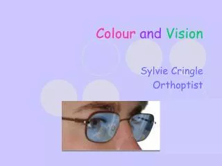
Colour Vision IIThe post receptoral basis of colour vision Prof. Kathy T. Mullen McGill Vision Research (H4.14) Dept. of Ophthalmologykathy.mullen@mcgill.ca 29th Sept 2005
Summary 1. Revision: cone types, the principles of trichromacy, univariance, and tests for the inherited color vision deficiencies 2. Connection of cones to retinal neurons: cone opponency 3. Cells types for RG, BY and Ach vision 4. Testing of RG, BY & Ach vision: 1) Farnsworth Munsell 2) Monitor displays and selective color vision tests 5. Examples from Optic Neuritis & Phototoxicity 6. Kollners Rule 29th Sept 2005
Spectral sensitivities of L, M & S cones Long Medium Log relative sensitivity Short Wavelength (nm)
Principle of Univariance • The response of a photoreceptor to any wavelength can be matched to any other wavelength simply by adjusting the relative intensities of the two stimuli Therefore: any single receptor type is colour blind
Principle of Trichromacy • Mixing together three coloured lights in suitable proportions enables us to make an exact match to any other colour • The 3 mixing lights are called ‘primaries’ • The match is called ‘metameric’ - meaning that identical colour sensations are produced even though the stimuli are physically different 3 mixing lights test light to be matched L1 + L2 + L3 L4
Colours with different wavelength distributions will look identical if they produce the same ratio of quantum catches in the L, M and S cone types
Trichromats • One of the three cone types is anomalous
Dichromats • One of the three cone types is missing
Mixing red and green lights to match yellow. A B C A and B. Green and red lights on the top are mixed by the subject to match the yellow light presented on the bottom. C. The red-green mixture perfectly matches the yellow. The same match as it appears to a deuteranomalous observer.
Ishihara test for RG color blindness 45 or spots 29 or spots 56 in both 6 or spots http://www.toledo-bend.com/colorblind/Ishihara http://www.vischeck.com/daltonize/
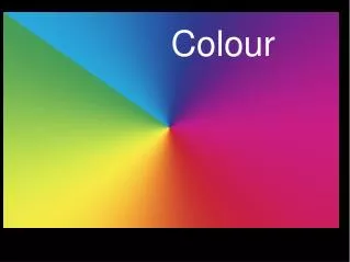
How is colour coded? • Each colour produces a unique pattern of relative activities in the three cone types
Connections of cones to retinal neurons http://webvision.med.utah.edu/index.html
Cones connect via retinal neurons into excitatory and inhibitory subgroups - - - - + - - http://webvision.med.utah.edu/index.html
Retinal cells Magnocellular (M) Parvocellular (P)
L/M (red-green) cone-opponency: P cells of retina & LGN S cone-opponency: bistratified ganglion cell & K cells of LGN Luminance (black & white): P cells and M cells. Neural pathways for color vision: How do we test these pathways? Farnsworth Munsell 100 hue or Panel D15 Electronic displays & computer graphics
Farnsworth Munsell 100 Hue Inherited and acquired color vision deficiencies Red-green, blue-yellow and non-specific deficiencies Show axial effects D15
Farnsworth Munsell 100 hue Protan Deutan Tritan
Show over heads for: F-M 100 hue in Optic Neuritis F-M 100 hue after intense light exposure
A pattern with colors that activate only the S/L-M (‘blue-yellow’) cone opponent process A pattern with colors that activate only the L/M (‘red-green’ cone opponent process
Contrast sensitivity of red/green and luminance gratings luminance red/green
Loss of colour and luminance contrast sensitivity with multiple sclerosis and optic neuritis Threshold RG BY Ach Patrick Flanagan and Connie Markulev Ophthalmic and Physiological Optics Volume 25 Issue 1 Page 57 - January 2005
Kollner’s Rule (1912) Lesions of the outer retinal layers affect blue yellow vision, lesions of the inner layers and optic nerve affect red-green vision Updated version S cones are physiologically vulnerable and so are more likely to be damaged by receptoral lesions than are L or M cones Post receptoral lesions are more likely to affect both types of cone opponent neuron: red-green and blue-yellow.
Conditions quoted as having tritan (BY) defects appearing first: Damage due to high light exposure Glaucoma Retinal detachment Pigmentary degeneration Myopic retinal degeneration ARMD Chorioretinitis Retinal vascular occlusion Diabetic retinopathy Papilledema Drugs: oral contraceptives, chloroquine S cones are genetically robust but vulnerable physiologically
Conditions quoted as having RG defects, but BY defects may also occur: Lesions of optic nerve/pathway Retrobulbar neuritis Leber’s optic atrophy Compressive lesions of the optic tract Progressive cone degeneration L and M cones are physiologically robust but genetically vulnerable