Download
1 / 22
250 likes | 504 Views
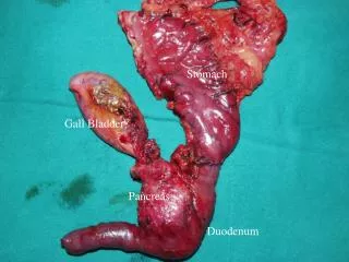
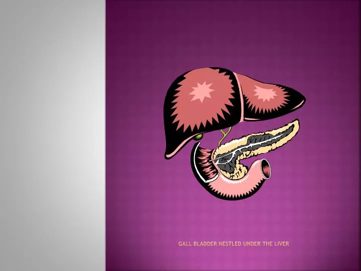
Gall bladder nestled under the liver. Gall Bladder. Location: under the liver Description: a sac-like organ located on the inferior surface of the liver Mucosa of the GB wall absorbs water and electrolytes resulting in a high concentration of bile salts, bile pigments and cholesterol.

E N D
Gall Bladder • Location: under the liver • Description: a sac-like organ located on the inferior surface of the liver • Mucosa of the GB wall absorbs water and electrolytes resulting in a high concentration of bile salts, bile pigments and cholesterol. • Primary purpose of the GB is to store and concentrate bile (90 mL)
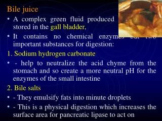
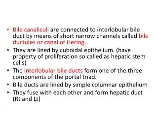
The path of bile • The cystic duct connects the gallbladder to the hepatic duct and they merge to form the common bile duct. • The sphincter of Oddi is at the distal end of the common bile duct and controls the flow of bile into the duodenum. • The bile secretions that empty from the common bile duct into the duodenum are necessary for digestion.
Cholecystitis • An inflammation of the gallbladder. • Remember back to inflammation and what happens within the body when that occurs.
Inflammatory response “The body’s celllular response to injury, infection or irritation. A protective vascular reaction that delivers fluid, blood products and nutrients to an area of injury. The process neutralizes and eliminates pathogens or dead (necrotic) tissues and establishes a means of repairing body cells and tissues.” Perry and Potter, p. 646
Signs and symptoms • Severe and steady pain in the upper right part of your abdomen. • Pain worsens when you inhale deeply. (Murphy’s sign) • Pain that radiates from your abdomen to your right shoulder or back. • Tenderness over your abdomen when touched • Sweating • N/V • Anorexia • Fever, chills, abdominal bloating
Causes • Gallstones • Injury • Infection • tumor
Risk factors • Gallstones • Cholesterol stones • Pigmented stones • Long labor • Traumatic injury • Diabetes
complications • Gallbladder distention • Infection • Tissue death • perforation
Questions to ask • When did you first begin experiencing symptoms? • Have you had bouts of pain similar to this before? • Do you have a fever? • Have your symptoms been continuous or occasional? • What improves your symptoms? • What makes them worse?
Tests and diagnosis • Blood tests • CBC • Hyperbilirubinemia • Elevated Erythrocyte sedimentation rate (ESR) • E-lytes • Alkaline phosphatase • Liver Function Tests (LFTs) • Imaging tests • Hepatobiliary iminodiacetic acid (HIDA) scan (aka cholescintigraphy, hepatobiliary scan)
Therapeutic Management • Oral dissolution therapy with ursodeoxycholic acid • Extracorporeal shock wave lithotripsy • ERCP = Endoscopic retrograde cholangiopancreatography • Laparoscopic cholecystectomy • Cholecystectomy
Priority Nursing diagnoses • Acute pain • Risk for impaired gas exchange related to pain and ineffective inspiratory effort • Imbalanced nutrition: less than body requirements related to nausea, vomiting and anorexia • Anxiety related to lack of knowledge about disease process and treatment measures
Interventions • Implement comfort measures • Provide education regarding diagnostic tests and disease process • Maintain NPO and Institute IV therapy as ordered • Nutrition counseling • Weight loss (3 Fs) • Monitor fluids and e-lytes
Medication Therapy • Symptomatic treatment of pain and nausea with analgesics and antiemetics • Meperidine (Demerol) is the preferred opioid analgesic because Morphine can cause spasms. • Cholestyramine (Questran) is used for severe cases of pruritus: Binds the bile salts to hasten excretion through the feces. • Chenodeoxycholic acid (CDCA) and urodoxycholic acid (UDCA) are oral dissolution medications
cholecystitis • Description: an acute or chronic disorder, most often caused by gallstones obstructing the cystic duct resulting in distention and inflammation of the gallbladder.
Etiology • Most commonly caused by gallstones blocking the cystic or common bile duct. • A small percentage of clients develop acalculous cholecystitis precipitated by trauma, prolonged hyperalimentation, fasting or surgery.
Assessment • Clinical manifestations include all those identified with cholelithiasis • Fever leukocytosis, elevation of serum bilirubin(possible jaundice), elevation of alkaline phosphatase, and elevation of amylase if pancreatic ducts are involved. • Abdominal guarding, rigidity, and rebound tenderness suggest peritoneal involvement.
Therapeutic management • NPO • IV hydration • Opioids for pain control • IV antibiotics • Surgical intervention is postponed until the acute infectious process has subsided.
Surgical interventions • Laproscopic cholecystectomy • Cholecystectomy • Cholecystectomy with T-tube placement
Postoperative care • Prevent infection • Control pain • Prevent pulmonary complications • Maintain T-tube is needed • Below the incision • Assess drainage and record amount • Assess skin at insertion site • Report bile drainage in excess of 500 mL after 3 days • T-tube is removed when drainage has subsided and stools have returned to a normal color
Postoperative care • Maintain NPO status as ordered • Advance diet slowly; low fat diet • Monitor bowel sounds • Encourage ambulation to promote peristalsis • Prevent DVTs • Provide general postoperative instructions: • Wound care • Analgesia • Diet • Signs of infection