Download

1 / 34
350 likes | 652 Views
Case presentation in infection disease 報告人: I2 李宗穎 日期: Jan.16, 2006. Chief Compliant. Female 22-year-old American Multiple ulcerated skin lesions on her face and extremities. Present illness. According to the statement of the patient, she was relatively healthy previously.

E N D
Case presentation in infection disease 報告人:I2 李宗穎 日期:Jan.16, 2006
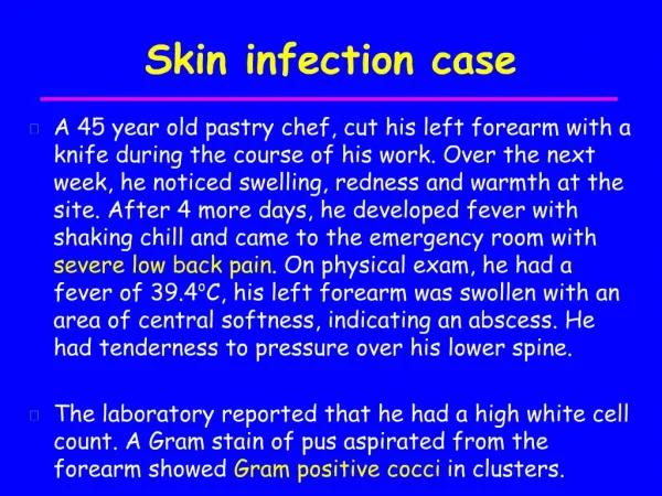
Chief Compliant • Female • 22-year-old • American • Multiple ulcerated skin lesions on her face and extremities.
Present illness • According to the statement of the patient, she was relatively healthy previously. • She went to Kenya for vacation 2 weeks ago. • She returned to Boston after a vacation. Multiple ulcerated skin lesion on her face and extremities was noted. • Due to persistent symptoms, she visited a local emergency room for help.
A Giemsa-stained smear showed monocytes containing small, oval, nonmotile parasitic forms measuring 2 to 3 μm in length.
Skin test • A skin test was performed and was strongly positive for a hemoflagellate (血液鞭毛蟲)
Questions 1,2,3 • Which infection does this patient have? What is the name of the hemoflagellate causing her infection? • Name the three species belonging to this complex. • Which vector is responsible for the transmission of this infection?
Hemoflagellates • 寄生於血液及組織內,屬於錐蟲科(Trypanosomatidae). • 須有無脊椎動物和脊椎動物兩種宿主,交替生長,才能完成發育。 • 錐蟲屬(Trypanosoma)及利什曼屬(Leishmania),對人類關係最密切。
Leishmania • 具a)無鞭毛期(amastigote)→於哺乳類中找到 b)前鞭毛期(promastigote)→只存於昆蟲媒介體內 • 全由白蛉(sandflies,沙繩)為媒介 a)只有雌蠅才產卵吸血,才具傳染能力 b)白蛉藉叮咬將promastigote注入到人體→被巨噬細胞吞入後發育為amastigote而繁殖 c)許多種哺乳動物都為其貯存宿主,如狐貍、貓、狗及囓齒類。
Etiology Several species of Leishmania are pathogenic for man: • L. donovani causes visceral leishmaniasis (Kala-azar, black disease, dumdum fever); • L. tropica (L. t. major, L. t. minor and L. ethiopica) cause cutaneous leishmaniasis (oriental sore, Delhi ulcer, Aleppo, Delhi or Baghdad boil); • L. braziliensis (also, L. mexicana and L. peruviana) are etiologic agents of mucocutaneous leishmaniasis (espundia, Uta, chiclero ulcer).
Cutaneous and visceral leishmaniasis also are endemic in parts of Eritrea, but the causative species have not been well established. • "L. infantum" and "L. chagasi" are synonymous. • L. tropica also causes leishmaniasis recidivans and viscerotropic leishmaniasis. • L. major-like organisms also cause New World cutaneous leishmaniasis. • The cutaneous leishmaniasis syndrome caused by this species is called.
Symptoms- Visceral leishmaniasis • Rapidly eliminated from the site of infection, hence there is rarely a local lesion • They are localized and multiply in the mononuclear phagocytic cells of spleen, liver, lymph nodes, bone marrow, intestinal mucosa and other organs. • One to four months after infection, there is occurrence of fever, with a daily rise to 102-104 degrees F, accompanied by chills and sweating.
The spleen and liver progressively become enlarged • With progression of the diseases, skin develops hyperpigmented granulomatous areas (kala-azar means black disease). • Untreated disease results in death.
Symptoms- Cutaneous leishmaniasis • Papule, 1-2 weeks (or as long as 1-2 months) after the bite. • The papule gradually grows to form a relatively painless ulcer. • The center of the ulcer encrusts while satellite papules develop at the periphery. • The ulcer heals in 2-10 months, even if untreated but leaves a disfiguring scar. • The disease may disseminate in the case of depressed immune function.
Ulcerative skin lesions with raised outer borders on the arm of a patient with New World (American) cutaneous leishmaniasis acquired in Costa Rica. (Photograph courtesy of Dr. A. Wright.) • People in Kabul, Afghanistan, infected with Leishmania tropica, standing in line for hours on a bitterly cold day in February 1997 at a treatment center for cutaneous leishmaniasis. [Photograph courtesy of Dr. R. Ashford and reprinted with permission from Elsevier Science (Lancet 354:1193, 1999).]
Symptoms-Mucocutaneous leishmaniasis • Initial- the same as those of cutaneous leishmaniasis, except that in this disease the organism can metastasize and the lesions spread to mucoid (oral, pharyngeal and nasal) tissues and lead to their destruction and hence severe deformity .
Questions 4,5 • Which stage of this parasite is the infective form? • Which stage of this parasite is seen in human blood smears?
Questions 6 • What is the skin test used on the patient? How is this test useful in screening the population in areas of endemic infection?
Montenegro test • A skin test with leishmanin (Montenegro test) is negative during active kala azar, but later becomes positive (after 6 to 12 months). • The Montenegro test reflects the suppressed cellular immunity during infection. There is a specific anergy for Leishmania parasites during active disease.
This test is mainly of epidemiological value. • To perform the test 0.1 ml is injected intradermally and the local reaction read after 48 hours(>5 mm induration = positive). • A positive test eliminates the existence of active kala azar. • Cutaneous leishmaniasis produces a positive Montenegro test.
Questions 7 • Which treatment is recommended for this infection?
Treatment • Most cases of cutaneous leishmaniasis heal within several weeks to 3 years without treatment. • Systemic therapy is indicated for infections caused by organisms in the L braziliensis species complex; →prevent mucosal leishmaniasis; and to decrease the morbidity caused by these generally chronic skin lesions.
For cutaneous L braziliensis infection, the CDC recommends treatment with sodium stibogluconate, 20 mg/kg/d intravenously for 20 days, because the cure rate has been only 64% using a lower dosage (10 to 13 mg/kg/d). • Common side effects include increases in hepatocellular enzyme levels, pancytopenia, and ECG changes consisting of T-wave inversion and prolongation of the QT interval.
Questions 8 • How is the diagnosis of this infection made?
Diagnosis • History of exposure to sandfies • Symotoms • Giemsa-stained slides of the tissue→most commonly used to visualize the parasite. • In vitro culture of infected tissue →unsuccessful because of contamination of the skin with yeast before the biopsy sample was taken • A skin test (delayed hypersensitivity: Montenegro test) • Detection of anti-leishmanial antibodies by immuno-fluorescence
Three examples of Leishmania spp. amastigotes from a skin ulcer. Note each amastigote contains a nucleus and the bar-shaped kinetoplast. These preparations have been stained with Giemsa stain.
Leishmania tropica amastigotes from an impression smear of a biopsy specimen from a skin lesion • In A, an intact macrophage is practically filled with amastigotes (arrows), several of which have a clearly visible nucleus and kinetoplast; in B, amastigotes are being freed from a rupturing macrophage.
Differential diagnosis Differential diagnosis includes • ulcers due to mycobacteria, • cutaneous diphtheria, 白喉 • tertiary syphilis, • yaws, • cutaneous carcinoma and deep or subcutaneous mycosis. • Acid fast bacilli can be made visible using the method of Ziehl-Neelsen. • Field sore (cutaneous diphtheria) and tropical ulcers (fusobacteria + Borrelia) are painful, particularly in the early phase.