Download
1 / 20
220 likes | 260 Views
Learn about different types of pathogens, how they cause disease, and the categories they fall into, such as bacteria, viruses, fungi, and more. Understand the differences between Gram-positive and Gram-negative bacteria and how they can affect health outcomes.

E N D
Pathogens By C. Kohn Agricultural Sciences Waterford, WI
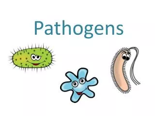
Pathogens • Any organism that is capable of causing a disease is called a pathogen. • Most pathogens are microorganisms (bacterium, virus, or fungus) but most microorganisms do NOT cause disease. • Many microorganisms even provide some protection from infectious pathogens by slowing their growth through competition. • In order to cause disease a pathogen must be able to get entrance into a host (the affected organism), adhere to the host’s tissue, and cause damage. • A pathogen most commonly gains entrance into an animal via the mucus membranes, including the mouth, eyes, nostrils, and genitals. • Cuts or openings in the skin can also lead to infection. Source: http://www.scq.ubc.ca/mucosal-immunity-and-vaccines/
Pathogens are Specific • Most pathogens attack a specific kind of tissue. • While a pathogen can invade the tissue where they gained entrance into the host, most often a pathogen focuses on attacking a specific kind of tissue in the host (such as respiratory cells, intestinal tissue, or other specific kinds of cells). • While the growth and reproduction of a pathogen can cause problems inside the host’s body, damage is more often due to the production of toxins or destructive enzymes by the pathogen. • These toxins or enzymes are often used to enable the pathogen to further invade the host’s tissues and/or to more easily acquire energy or nutrition from the host. • For example, some ‘flesh-eating’ diseases produce enzymes that break down tissue and dissolve fibrin blot clots in order to enable the pathogen to invade even more tissue in the host. Source: onlineimmunology.blogspot.com
Categories of Pathogens • There are six major kinds of pathogens that can cause infectious disease: • Bacteria: single-celled organisms that lack cellular organelles and divide by fission (splitting in two). • Viruses: non-living nucleic acid surrounded by a protein coat that uses living cells to reproduce. • Fungi: eukaryotic (has organelles) organisms that reproduce by forming spores, can be unicellular or multicellular, and are common decomposers. • Protozoa: single-celled eukaryotic organisms that are mobile and feed off other organisms. • Helminths (worms): simple multi-celled invertebrates that often have multi-staged reproductive cycles. • Prions: non-living infectious proteins that most commonly affect the nervous system of the host. Source: www.riversideonline.com
Bacteria (Prokaryotes) • Bacteria are unicellular (single-celled) organisms. • Bacteria are prokaryotes, meaning they lack a nuclei, mitochondria, or other cellular organelles. (Conversely, eukaryotes have organelles like a nuclei or mitochondria.) • They have circular, double-stranded DNA. • They also have small additional ‘packets’ of DNA called plasmids. • Most bacteria reproduce by growing and then dividing into two cells in a process called binary fission. • Bacteria are typically classified by their shape. • Bacteria are most commonly classified as either bacillus (rodshaped), coccus (spherical), or spirillum (helical rods). • Bacteria can also be classified by how they obtain their energy. • Some bacteria are photosynthetic, some oxidize inorganic compounds, and some break down organic compounds (such as sugar and amino acids). • Bacteria can also be classified as aerobes (need oxygen), anaerobes (can only live in the absence of oxygen) or facultative anaerobes (can live with or without oxygen). Source: http://evolution.berkeley.edu/evolibrary/images/endosymbiosis/cells.gif
Gram Neg vs. Gram Pos • In regards to disease, bacteria are most commonly classified as Gram Positive or Gram Negative based on the presence or absence of an outer cell membrane. • Both kinds of bacteria are very similar internally. • The main difference between a gram negative and a gram positive bacteria is based on their cell walls. • Gram positive and gram negative bacteria can be identified using a laboratory stain. • When a gram stain is applied, gram positive bacteria turn purple and gram negative bacteria turn red. Source: http://www.dbriers.com/tutorials/wp-content/uploads/2012/12/GramPositiveNegative21.jpg
Gram Positive Bacteria • A gram positive bacteria has a cell wall made mainly of peptidoglycan. • Peptidoglycan is a mesh-like substance in the cell wall and is similar to that of the exoskeleton of an insect. • Because the peptidoglycan cell wall is mesh-like, this means that substances can diffuse across the membrane and enter the inside of the bacterial cell. • This makes gram positive bacteria susceptible to most antibiotics, and this makes it easier to treat a gram positive bacterial infection than it is to treat a gram negative bacterial infection. • Peptidoglycan can also be broken down by lysozyme enzymes produced by animal cells. Source: studydroid.com
Gram Negative Bacteria • Gram negative bacteria have an extra layer of protection due to the presence of an outer membrane on the outside of their cell wall. • This outer membrane is like a stiff canvas sack and blocks larger molecules including antibiotics and lysozymes. • The outer membrane is like a bulletproof shield for gram negative bacteria, repelling most molecules that would otherwise harm the bacterial cell. • The outer membrane also protects gram negative from drying and from harsh environments including the stomach acid of animals and engulfment by white blood cells. • Finally, the outer membrane can enable some species of gram negative bacteria to adhere (or ‘stick’) to the cells of their hosts to increase their likelihood of invasion and infection. Source: http://biology-forums.com/index.php?action=gallery;sa=view;id=752
Toxins • Gram negative bacteria contain endotoxins in their cell wall and outer membrane. • Endotoxins are toxins found inside bacterial cells and are mostly only released if the cell if broken down. • This is different from an exotoxin, which is a toxin released by a bacterial cell while it is still alive. • Exotoxins are much more common in gram positive bacteria, whereas endotoxins are more common in gram negative bacteria. • If the outer membrane and cell wall of gram negative bacteria are broken down, these endotoxins will be released into the body of the host. • Endotoxins are very resilient and can remain intact even after 30 minutes of boiling temperatures. • These endotoxins cause an inflammatory response in animal hosts. Source: www.ib.bioninja.com.au
Septic Shock • The inflammatory response due to the presence of toxins from bacteria can lead to septic shock and even death. • When a toxin is sensed by an animal’s body, it causes a systemic (body-wide) inflammatory response. • This means that all the blood vessels in the body expand. • As vasodilation (expansion of the blood vessels) occurs, the blood pressure of the animal drops. This drop in blood pressure is known as hypotension. • The heart will weaken as it works harder to compensate for the hypotension. • As a result of this hypotension, organs will not receive adequate oxygen or nutrients due to impaired blood flow (hypoperfusion), and organ systems will begin to shut down. • The kidneys will be unable to eliminate waste, allowing it to accumulate in the blood. • The respiratory system will begin to fail, resulting in even less oxygen flow to the body’s organs and an increased rate of organ shut-down. • When an infection causes life-threatening low blood pressure, this is known as septic shock.
Stages of Septic Shock Infection & Bacteremia • Septic shock has the following stages: • Infection: presence of bacteria in what is normally sterile bodily tissue. • Bacteremia: presence of bacteria in the blood. • Systemic Inflammatory Response (SIRS): when blood vessel dilate (vasodilation) due to bacteremia, causing hypotension (drop in blood pressure) and hypoperfusion (lack of blood flow through an organ). • Sepsis: when inflammatory responses occur in tissues that are remote from the infection. • Sepsis becomes septic shock if the systemic inflammatory response leads to dangerously low blood pressure, and organ systems begin to fail as a result. SIRS Sepsis SepticShock Death
Viruses • The second category of disease-causing pathogens are viruses. • A virus consists of genetic material surrounded by a protein coat. • Viral genomes can be double or single stranded and can be DNA or RNA • Viruses are non-living. They cannot reproduce on their own and they do not metabolize food for energy. • Because a virus is not alive, it will not respond to an antibiotic. • To reproduce, a virus must hijack a cell and manipulate the cell so that it produces viral proteins instead of its normal cellular proteins. • To do this, the virus inserts its genome into the host cell, and forces the cell to reproduce its own genome. • The cell then makes mRNA and tRNA in order to produce more viral proteins. • The cell assembles the viral proteins and genomes into new viruses. • These newly-assembled viruses are released from the cell and then infect other cells, repeating the process over and over. Source: learningon.theloop.school.nz
Retroviruses • Retroviruses are a kind of virus with the ability to insert their own genetic material into the genome of a cell. • Retroviruses have a unique enzyme called reverse transcriptase that allows them to copy their RNA into the cell’s genome. • As the host’s cells divide, they reproduce the viral DNA, making retroviruses difficult to eliminate from a host. • Retroviruses also tend to have long latent periods (the time between infection and the exhibition of symptoms) which means that the disease often goes unnoticed and can more easily spread. • HIV is an example of a retrovirus. • All viruses cause their respective diseases by interrupting normal cellular function. • Some viruses use their own proteins to stop the creation of the host cell’s proteins. • Some viruses cause the cell membrane to break open and rupture. • Some viral proteins are toxins. • The presence of some proteins causes the host’s own immune system to attack and destroy its own cells to eliminate the virus.
Fungi • The third category of pathogens are fungi. • Fungi, like animals and plants, are classified as their own kingdom of life. • Like animals and plants, fungi are eukaryotic, meaning they have cellular organelle. • Fungi can be either unicellular (such as yeast) or multicellular (such as mushrooms). • With bacteria, fungi are the main decomposers in the environment. • Ringworm in livestock is a well-known example of a disease caused by a fungal pathogen. Source: http://www.scabies-killer.com/images/ringworm.jpg
Protozoa • Protozoa are the fourth category of pathogens. • Protozoa are single-celled eukaryotes (they have cellular organelles). • The amoebas and paramecium are common examples. • Protozoa lack cell walls, which make them flexible and capable of quick movements. • Protozoa often invade the tissue of their hosts, causing tissue erosion and degradation. • Other protozoa, including Giardia, cause infection of the large intestine, causing it to swell which prevents nutrient absorption and causes diarrhea, gas, and cramping. • Malaria is caused by the Plasmodium protozoa; when Plasmodium gets into the blood, it destroys red blood cells and causes anemia, alternating fever & chills, exhaustion, and often death. Source: http://thumbs.dreamstime.com/z/protozoa-22943129.jpg
Helminths • Helminths, or parasitic worms, comprise the fifth category of pathogens. • Helminths are multicellular eukaryotes with tube-like bodies. • There are three main classes of helminthes: nematodes (roundworms), cestodes (tapeworms), and trematodes (flukes). • Helminths are unique because they do not proliferate inside their hosts; their offspring will usually be passed in fecal matter from animal hosts so that they can infect other animals. • Most helminths develop slowly inside their hosts, and usually symptoms are mild and have a slow onset. • Helminths can affect their hosts in a variety of ways. • Both adults and larva can cause diseases depending on the species. • The severity of the symptoms depends on the concentration of helminths inside the host. • Helminths affect the host’s tissue in a number of ways, but typically they cause disruption either by physically disrupting the tissue of the host or by taking nutrients from the host’s body. Source: http://bingsti.ru/styled/files/044004380441-1.jpg
Examples of Helminths • Examples of helminth diseases include the following: • Hookworms are a kind of helminth and cause anemia (lack of red blood cells) and malnutrition. • Some helminths burrow into the skin or eyes causing itching, infection, and inflammation. • Cysticercosis is caused by a pork tapeworm and causes bumps to develop in the skin and muscles as well as neurological problems. • Echinococcus tapeworms cause liver failure, lung disease, and brain abnormalities. Source: http://www.poultryhub.org/wp-content/uploads/2012/05/Helminths-Ascaridia-galli-in-SI.jpg
Prions • Prions are the most-recently discovered class of pathogens. • Prions are infectious proteins. They are not alive and are not a kind of living species. • Prions affect their host by causing abnormal folding of the host’s proteins. • Like a key for a lock, the function of a protein depends on its shape. • When a prion alters the shape of a protein, it changes its function and makes it useless (much like if you bent a key at an angle, it would not work in a lock). • The abnormal folding or proteins leads to tissue loss in the brain of the host, leading to literal holes in the brain. • Bovine Spongiform Encephalopathy (BSE, or Mad Cow), Chronic Wasting Disease (CWD, common in deer and elk), and scrapie (common in sheep) are common forms of animal prion diseases. • Humans prion diseases include Creutzfeldt-Jakob Disease (CJD) and Kuru.
Prion Diseases • Prion diseases are most commonly spread through ingestion of infected materials. • For example, scrapie and mad cow disease (below) were transmitted to animals when they were fed rendered animal protein supplements from previously-infected animals. • Kuru was spread among the Fore people of New Guinea because of a practice of ritualized cannibalism. • CWD seems to be spread by saliva, urine, and feces and may have a correlation to populations of deer and elk that have high concentrations around a feeding area. • As of this time, there is no known treatment for any prion diseases. Source: quizlet.com Source: news.bbcimg.co.uk/media/images
Works Cited • http://micro.digitalproteus.com/morphology2.php • http://www.path.cam.ac.uk/~schisto/general_parasitology/parasitology_general_pathology.html • http://www.merckmanuals.com/home/infections/bacteremia_sepsis_and_septic_shock/sepsis_and_septic_shock.html • http://www.cdc.gov/ncidod/dvrd/prions/ • http://www.ninds.nih.gov/disorders/kuru/kuru.htm • http://www.cwd-info.org/index.php/fuseaction/about.faqDetail/ID/209ea1b39c93f85dde9a5a4261400ea2 • https://science.education.nih.gov/supplements/nih1/diseases/guide/understanding1.htm • https://www.softchalk.com/lessonchallenge09/lesson/ImmuneSystems/BloodLymphaticandImmuneSystems_print.html