Download
1 / 72
1.17k likes | 3.31k Views
LASIK COMPLICATIONS . Eric E. Polk, OD, FAAO. LASIK Complications. Objective The student will be familiar with the most common intra-operative and post-operative complications of LASIK surgery. LASIK Complications. Most common problems Blurred vision: Under or over correction

E N D
LASIK COMPLICATIONS Eric E. Polk, OD, FAAO
LASIK Complications • Objective • The student will be familiar with the most common intra-operative and post-operative complications of LASIK surgery
LASIK Complications • Most common problems • Blurred vision: Under or over correction • Glare, halos and night vision problems • Dry eye • Microstriae • Infrequent complications • Diffuse lamellar keratitis (DLK) • Epithelial Ingrowth • Flap slip • Irregular ablation: Ghosting image, double image • Staph marginal infiltrates • Pressure induced stromalkeratitis • Intraoperative complications • Corneal abrasion • Irregularly thin flap • Less frequent/ more devastating • Infection • Keratectasia • Flap complications: Buttonhole flap, free flap, irregular flap • Decentered ablation • Programming Error
Blurred Vision • No promise of 20/20 vision • 89% 20/20 Vision • 3-4% undergo an enhancement surgery • Eyes may over or under correct. • Induced astigmatism • Regression of vision • Higher Rx: Increased risk: More variability • Biomechanics of cornea • Some variables you can consider • Curvature, thickness, high order aberrations • Other variables are not easily measured • Hydration of cornea, density of cornea, cell migration following surgery
Custom Wavefront= Perfect Vision? • Lipshitz, Isaac. 34 Challenges to Meet Before Excimer Laser Technology Can Achieve Super VisionJ Refract Surg. 2002 Nov-Dec;18(6):740-3. • Changes in wavefront with age • Changes in wavefront with accommodation • Biomechanical differences among corneas • Changes in tear film • Corneal epithelium wound healing • Challenges met • Pupil Tracking • Iris Registration
Treatment • Enhancement surgery • Wait 4-12 months for RX stability • Two RX checks • Lower RX: Faster stabilization • Hyperopes take longer • Enhance by • Lifting flap • Cutting new flap • PRK enhancement over LASIK flap for older flaps • Risk involved with flap lift enhancement surgery • Thinner cornea: keratectasia • Epithelial ingrowth
Patient Satisfaction • Patient’s perception of successful surgery is different for each person • 3.0% will seek out enhancement surgery • Risk factors for unsatisfactory outcome • High myopes/astigmatism • Hyperopia • Age/Monovision • Patients taking antidepressant or medications for anxiety • Morse JS, et al. Role of depressive symptoms in patient satisfaction with visual quality after LASIK. J Cataract Refract Surg. 2009 Feb: 35(2): 341-6
Night Vision ProblemsGlare and Halo • Most common side effect: Possible permanent complication • Risk factors • High RX • High Astigmatism • treatment area is smaller and cannot blend as well • Large pupils: undilated • Average pupil 6.0. Range 3-9.5 mm • Small optical zones • Standard laser ablation
Treatment • Time: Glare and halo most common after surgery (first month) then fades with time, up to six months • Miotics: Alphagan, Pilocarpine. • Custom wavefront enhancement • Correct spherical aberration
Dry Eye • Dry eye is one of the most common side effects of surgery • Season • Winter • Work Environment • Hospitals • Geography • Desert
FDA hearing on Lasik complaints reveals fury and despairAgency conducts study to identify who has bad outcomes and whyBy LAURAN NEERGAARDAssociated Press • Published on: 04/25/08 WASHINGTON (AP) — Patients harmed by Lasik eye surgery alternated between fury and despair Friday as they told federal health officials of suffering years of eye pain, blurred or double vision — even of people driven to suicide. • "Too many Americans have been harmed by this procedure and it's about time this message was heard," said David Shell of Washington, D.C., who had Lasik in 1998 and says he has "not experienced a moment of crisp, good quality vision since."
Pre-Treat Dry Eye Patients • Patients with pre-operative dry eye may develop significant dryness following surgery • SPK may alter wavefront measurement before surgery • SPK may alter pre-operative refraction
Pre-operative Dry Eye Treatment • Mild • Artificial tears • Moderate • Artificial tears • Ointment • Restasis • Severe • Preservative free tears • Gels • Restasis • Mild steroids (Lotemax) • Punctal plugs • Flax seed or fish oil (omega 3 fatty acids)
What causes Dry Eye after Surgery? • Severed corneal nerves • Reduced corneal sensation (corneal hypoesthesia) • Decreased blink rate • Decreased reflexive tear production • Neural feedback to lacrimal gland
Benitez-del-Castillo J, et al. Decrease in tear secretion and corneal sensitivity after LASIK. Cornea 2001;20:1:30-32 • Corneal sensitivity measured following LASIK • 20% of baseline at one month • 50% at three months • 90% at six months • Normal values at nine months
Results of Study • Decreased blink rate. • Greater periods of unprotected time for the surface after the tear film breaks • Values were worse in long term contact lens wearers
Etiology: Goblet Cell Damage • Prolonged microkeratome pressure can damage conjunctival goblet cells • Unstable mucin layer after LASIK • Normal mucin layer is necessary to prevent evaporation of the tear film. • Patel S, et al. Corneal sensitivity and some properties of the tear film after LASIK; Journal of Refract Surg 2002: Mar-Apr; 18:2:113-23.
Etiology: Change in curvature • Change in corneal curvature • Myopic ablation • Flatter cornea • Hyperopic ablation • Steeper cornea • Decreased tear film coverage • Change in tear film distribution • Albeitz et al. Effect of LASIK for hyperopia on tear film and ocular surface. Journal of Refract Surg 2002:18;113-22.
Variables • LASIK effected more then PRK • Hyperopic ablations > Myopic ablations • High myopia > low myopia • Microkeratome > Intralase
Post-operative Dryness • Dry eye patients are often asymptomatic • Decreased or fluctuating vision may be only symptom • Decreased blink rate • Neurotrophic cornea • Less tear production
Treatment • Artificial tears • mild preservative or pres free • Gels or ointments • Punctal plugs: 30 day or permanent • Restasis • Fish oil • Omega 3 Fatty Acids
Epithelial Ingrowth • Proliferation of epithelial cells in the interface. • A progressive sheet of cells will grow under the flap once they are introduced. • Significant epithelial ingrowth can lead to the release of enzymes that may cause a flap melt.
Epithelial Ingrowth Presentation • More likely to occur after enhancement sx • Typically presents 3 weeks after surgery • May begin with a very subtle appearance • Epithelial cells are transparent • Leading edge of epithelial cells
Epithelial Ingrowth Leading Edge
Risk Factors for Epithelial Ingrowth • Rarely occurs after primary LASIK • Flap lift (enhancement surgery) • Intra-operative epithelial defects at the edge of the flap • Poor flap edge adhesion
Epithelial Ingrowth • Occurs in ~4% of enhancement surgeries • Bandage contact lenses decrease the risk of epithelial ingrowth
Epithelial Ingrowth • Less aggressive cases can be monitored • May cause erosion or fibrosis of the flap margin • Severe cases require flap lift and scrape • 50% chance of reoccurrence
Alternative Treatments for Epithelial Ingrowth • Baxter Tissue Sealing Glue • Tissuesealing.com • YAG Laser Ablation • Ayala MJ, et al. Treatment of LASIK interface epithelial ingrowth with neodymium YAG laser. Am J Ophthalmol. 2008 Apr;145(4):630-634. • Corneal sutures for epithelial ingrowth • Spanggord HM, et al . Flap suturing with proparacaine for recurrent eptihelialingrowthfollowin LASIK surgery.. J Cataract Refract Surg. 2005 May;31(5):916-21.
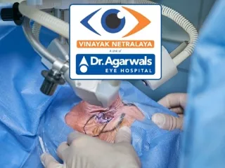
Post-LASIK Infection Devastating complication of LASIK surgery Can quickly cause degradation of flap tissue and loss of BCVA Incidence 0.1% of LASIK surgeries
Increased Incidence 2001 Survey: 1/2919 2004 Survey: 1/2131 2008 Survey: 1/1102 Infectious KeratitisFollowing Refractive Surgery:ASCRS 2008 Survey Results Solomon R, Donnenfeld ED, Azar DT, Holland EJ, et al. Infectious keratitis after laser in situ keratomileusis: results of an ASCRS survey. J Cataract Refract Surg. 2003;29:2001-2006.
Post-LASIK Infection Risk factors • Swimming pools, lakes, ponds, saunas, hot tubs • Dirty, dusty enviornments • Blepharitis • Meibomianitis • Health care providers • H-MRSA
Infectious Keratitis MRSA tend to cause the majority of infections at this time Mycobacteria is less of a concern then before Moxifloxacin and gatifloxacin cover the atypical mycobacteria
MRSA • Infections occurring the first week after surgery are more likely to be MRSA • After two weeks: Fungal, mycobacterial, nocardia • Patients at risk for MRSA • Healthcare workers • MRSA skin conditions • IV catheters • IDDM
Symptoms Pain, photophobia and redness Tearing Foreign body sensation Decreased vision Increase of glare and halos
Presentation of Infection • Anterior chamber reaction. • Epithelial defect over infiltrate. • Focal infiltrate with anterior and posterior extension from the interface.
Differential Diagnosis of Infection • DLK • Sterile Infiltrate • Surgical Debris • Microbial Keratitis • Bacterial • Mycobacterial • Fungal • Viral • Joshua Stein, MD • Ophthalmology Times
Microbial Keratitis following LASIK Lou Probst, MD
Treatment of LASIK Infectious Keratitis • Aggressive treatment of pre-operative blepharitis • If a infiltrate is identified within two weeks of LASIK/PRK surgery • Culture • Fortified Vancomicin • 4th Generation Fluoroquinolone • Discontinue steroid with open wound defect • Daily cornea checks
Treatment of Infectious Keratitis • Doxycycline to decrease collagen destruction • Loss of stromal tissue may occur quickly • Flap amputation • Joshua Stein, MD • Ophthalmology Times
Peripheral Keratitis • Staph. Marginal Infiltrates • Marginal Sterile Infiltrates
Etiology • Localized corneal hypersensitivity reaction to toxins produced by bacteria in eyelid margins. • Staphylococcus aureus most common • Also • Streptococcus pneumoniae and Pseudomonas aeruginosa • LASIK patients may be prone to this.
Staph Marginal Infiltrates • More common in patients with anterior and posterior blepharitis Dryeyezone.org
Staph Marginal Infiltrates • Patients are often asymptomatic • Does not effect vision • Sterile • No A/C Rxn • No Redness • Clear zone between infiltrate and limbus
Identification • Occur most often at lid margin and flap margin. • Occurs at the surface of the cornea, not the interface. • Sterile. • Secondary DLK may occur.
Treatment • Intensive topical corticosteroid. (Prednisilone 1%) • Lid Scrubs • Doxycyline 100 mg BID. • Antibiotic coverage. • Culture not necessary. • Close follow-up.