Download
1 / 54
540 likes | 662 Views
Neurology revision. 11 March 2014 Antony Thomas Consultant Neurologist. History. General Approach Is this neurological? If so where in the neuroaxis: central or peripheral? Above or below foramen magnum? Above or below the tentorium? What might be the nature of the problem?

E N D
Neurology revision 11 March 2014 Antony Thomas Consultant Neurologist
History • General Approach • Is this neurological? • If so where in the neuroaxis: central or peripheral? • Above or below foramen magnum? • Above or below the tentorium? • What might be the nature of the problem? • Differential diagnosis • Handedness
History Taking • Vital importance • Good listener • Focused • Lateral thinking • Anatomical and Pathological Diagnosis • Age / Occupation / Handedness • Temporal features of a symptom : 1.Onset 2.Progression 3.Duration 4.Recovery 5.Frequency • Weakness of one side of the body • Numbness of hands and legs
Direct Questions Pain Headache Facial, neck, back and limb pain Disturbance of consciousness Blackouts, faints, fits Altered sleep pattern Cognitive & affective dysfunction Memory, language Depression, irritability
Direct Questions Cranial Nerve symptoms • Loss of vision, blurring, diplopia • Hearing, sense of taste and smell • Facial muscle weakness • Vertigo, dizziness, giddiness • Bulbar muscles ( swallowing , articulation of speech)
Questions Limb symptoms • Difficulty lifting , gripping, fine finger movements, clumsiness • Gait disorder, leg weakness, stiffness, balance problems • Loss of sensation, altered sensation, numbness • Involuntary movements, incordination • Bladder, bowel, sexual dysfunction
Initial Impression • Gait • Facial Expression • Handshake • Speech • Arm swinging Positive symptom and negative symptom
History • Speed of onset • Instantaneous • Minutes • Hours • Days • Weeks/Months • Months/Years
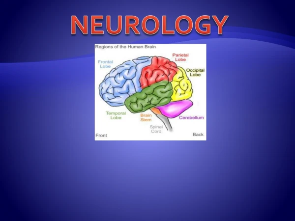
Anatomically lesions localised toWhere is the lesion? Meninges (Venus Sinus) Spinal fluid Cortex Subcortex (Basal Ganglia, Thalamus, Hypothalamus) Brain Stem (Midbrain, Pons & Medulla oblongata) Cerebellum Foramen magnum (Craniocervical Junction) Cranial Nerves Spinal Cord (ends at Lower border L1) Anterior Horn Cell Disorder Nerve root (Dorsal & Ventral) Plexus Peripheral Nerve Neuromuscular Junction Muscle
History • Instantaneous • “Electrical events” • Epilepsy • Myoclonic jerks • Neuralgic pain • Vascular events • Subarachnoid H’age (SAH) • Intracerebral H’age
History • Maximal over minutes • Vascular events • Migranous events • Maximal over Hours • Infective events • Inflammatory: GBS, Myelitis • Vascular: stroke • Vasculitic:GCA, Mononeuritis multiplex
History • Maximal over Days • Intoxication: Iatrogenic • Infection: HSV encephalitis, Meningitis • Inflammation: MS, GBS
History • Maximal over weeks/months • Brain tumours • Expanding unruptured aneurysms • Degenerative: CJD • Some polyneuropathies • Some myopathies: Steriod induced
History • Maximal over months/years • Neurodegenerative • Parkinson’s (PD) • Alzheimer’s • Cerebellar ataxias • Motor Neurone Disease (MND) • Most Neuropathies • Most myopathies
Single or multiple • Migraine • Epilepsy • TIA • Syncope • Trigeminal Neuralgia • Multiple Sclerosis (Relapsing Remitting)
Documenting Hx • No different in Neurology • Presenting complaint (PC) • Hx of the PC • Past Medical: Injuries, Psychiatric, Op, Arteriopath • Medication: Recreational use • Social/Employment: Driver, Smoker / Alcohol • Family Hx: Stroke, MND, PD, Dementias, Tremors, DM, MS
Common presenting complaints in Neurology • Funny turns • Seizures and LOC • Headaches • Dizziness & Vertigo • Confusion • Weakness of arms / legs • Abnormal movements • Loss of balance • Walking difficulties • Numbness and tingling, pins and needles • Visual failure, diplopia
HPC • As much detail as possible • When and where • Previous episodes • Witness accounts • Exacerbating and relieving factors • Treatments and changes to Rx • Associated symptoms
Recurrent attacks of LOC • Postures and manoeuvres • Drugs/Alcohol • Palpitations • Prodromal features • Post-ictal confusional states • Eye witness account • Treatments
Examination • Higher Mental Functions • Cranial Nerves • Motor • Sensory • Cerebellar • Gait • Sphincters • Skull and Spine • Neck stiffness • Neurocutaneous markers • General examination • Other systems
Higher Mental Functions • Appearance and behaviour • Mood and Affect • Thought form and content • Sensorium (GCS) and Cognition • Awareness • Sleep • Drowsiness • Stupor • Coma • Perceptual disturbances: Hallucinations • MMSE • Speech and Language
1. Olfactory 2. Optic 3. Occulomotor 4. Trochlear 5. Trigeminal 6. Abducens Smell Vision Elevate, depress and adduct, pup: constrict Depression, adduction, intorsion Face sensation, muscles of mastication Abduction Cranial nerves
7. Facial 8. Vestibulocochlear 9. Glossopharyngeal 10. Vagus 11. Spinal Accessory 12. Hypoglossal Muscles of facial expression, Anterior 2/3 tongue taste Hearing and balance Taste posterior 1/3 tongue, gag reflex Gag reflex, motor to soft palate, pharynx, larynx. Autonomic fibres to oesophagus, stomach, small intestine, heart, trachea, viscera Sternocleidomastoid, Trapezius Motor control tongue Cranial Nerves
Motor System • Bulk and nutrition • Wasting • Tone • Power • Reflexes • Babinski
DTR • 0 Absent • +/- Present with reinforcement • + Reduced • 2+ Normal • 3+ Increased Brisk Exaggerated • 4+ Pathologically brisk with clonus
Sensory System • Side to side • Proximal to distal • Pin prick • Touch • Vibration • Joint position sense • Romberg’s • Cortical sensation
Cerebellar signs • Intention Tremors • Titubation • Ataxia • Truncal ataxia • Dysdiachokinesis • Slurred speech and dysarthria • Hypotonia • Past pointing Dysmetria • Nystagmus • Tandem walking heel-toe walking • Rebound phenomenon • Pendular knee jerk • Hyporeflexia • Finger nose / Heel shin co-ordination (watch out for weakness)
Gait • Normal • Hemiplegic / Circumduction • Parkinsonian • Cerebellar • High stepping/ steppage or stamping • Waddling / Trendelenburg • Spastic • Scissor gait • Antalgic • Functional
Diagnostic tests • CSF analysis (LP) • EEG • Evoked Potentials • EMG • NCS • CT • MR • DAT • SPECT • Bloods
Typical Cerebrospinal Fluid Findings in Various Types of Meningitis Test Bacterial Viral Fungal Tubercular Opening pressure Elevated Usually normal Variable Variable WBC≥1,000 per mm3 <100 per mm3 Variable Variable Cell differential Predominance of Predominance of Predominance Predominance PMNs* lymphocytes†of lymphocytes of lymphocytes Protein Mild to marked Normal to elevated ElevatedElevated elevation CSF-to-serum glucose Normal to marked Usually normal Low Low ratio decrease CSF = cerebrospinal fluid; PMNs = polymorphonucleocytes. *—Lymphocytosis present 10 percent of the time. †—PMNs may predominate early in the course.
EEG • Encephalitis • Seizure Disorder • Encephalopathy • Anoxic brain injury • Degenerative conditions (CJD)
Trimodality EPs • Visual Evoked Responses • Brain Stem Auditory Evoked Response • Somatosensory EP
EMG/NCS • Muscle vs Motor Neuron • Demyelinative vs Axonal • Nerve root vs Plexopathy • Localisation of mononeuropathy • NMJ disorders: MG, LEMS • Entrapment Neuropathy
Neuropathy • Demyelinating • Slowed conduction • Preserved amplitude • Axonal • Reduced amplitude • Normal NCV
Neuroradiology • CT Head +/- contrast • MRI (MRA, MRV) • DWI (acute stroke) • PWI • FLAIR • MR Angiogram • PET/SPECT
Neurological Emergencies • Status Epilepticus • Coma • Traumatic Brain Injury (TBI) • Acute Stroke • Infections (Meningitis) • Subarachnoid Haemorrhage • Raised intracranial pressure Herniation • Acute Spinal cord compression • Acute Neuromuscular respiratory paralysis • Acute Visual loss • Delirium
Clinical scenario • 35 years old lady • 2/7 ago started with pins and needles in feet followed by difficulty walking then in the last 24 hours unable to hold a cup in her hands and could not get out of the bed • Past: Had diarrhoeal illness2 weeks ago. • O/E:-
O/E • Hypotonia • Faccid weakness • Areflexia • Bilateral Bell’s palsy • No UMN signs • Glove and stocking sensory disturbance • Diagnosis ??
GBS History Examination Flaccid weakness Hypotonia Hyporeflexia Cranial nerves involvement Respiratory muscle involvement Autonomic involvement Sensory disturbance No UMN signs
GBS Mortality rate 3 to 5 % Symmetric rapidly progressive, ascending, flaccid paralysis from a demyelinating poly radiculoneuropathy Post infective, post inflammatory 10% starts in ULs Progresses over the initial days up to 4 weeks Plateaux and then improves afterwards Proximal weakness Bells palsy in 50% Prior infection GIT/Resp
Diagnosis of GBS Classical history & findings Neurophysiology: Slowing of nerve conduction Serology: Campylobactor, CMV, EBV, HSV, Mycoplasma Antibodies: Anti GM1, Anti GQ1b CSF analysis: High protein with normal cells (Albumino-cytological dissociation) (? Neuro-imaging) Papilledema in GBS