Download
1 / 40
410 likes | 798 Views
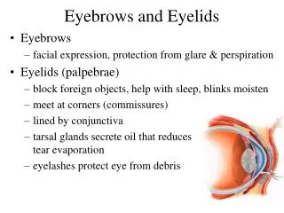
THE EYELIDS. THE EYELIDS The eyelids protect the eye from injury and excessive light. And prevent excessive dryness of the cornea and conjunctiva . Macroscopic anatomy : The eyelids are two movable mucocutaneous folds which act as shutters.

E N D
THE EYELIDS • The eyelids protect the eye from injury and excessive light. And prevent excessive dryness of the cornea and conjunctiva . Macroscopic anatomy : • The eyelids are two movable mucocutaneous folds which act as shutters. • The palpebral fissure is the space enclosed between the two lid margins when the lids are open. • In adults the palpebral fissure is 30 mm in length and 15 mm in width.
Position: the upper eyelid covers the upper 1/6 of the cornea (2mm). The lower eyelid is at the level of the limbus The eyelid is formed of 6 layers: Skin: very thin skin loosely attached to the underlying structures. Subcutaneous areolar layer: loose connective tissue containing no fat. Muscular layer: containing the Orbicularis oculi muscle. Submuscular layer: loose connective tissue containing the main blood vessels and nerves of the lid.
Tarsus: • the tarsal plate is a condensed fibrous tissue resembling cartilage, it acts as the skeleton of the lid. • The ends of the 2 tarsal plates are fixed to the orbital bones by the lateral and medial palpebral ligmants. Embeded in the upper tarsus are 20-30 vertically arranged meibomian glands. The lower tarsus contains 10-15 meibomian glands. Palpebral conjunctiva: • The conjunctive is very thin, vascular, and firmly adherent to the tarsus by fibrous bands
The muscles of the lid: • Orbicularis oculi muscle: the orbicularis muscle is the sphincter of the lid and has 4 portions. • Palpebral portion: it is the central part of the muscle and may be divided into presseptal and pretarsal parts. Action: simple closure of the lids as in blinking. It supports the lower lid in its place. • Orbital portion: Action: tight closure of the lids. • Horner's muscle (pars lacrimalis): a thin layer of muscle fibers arising from the posterior lacrimal crest and lacrimal fascia, when this muscle contracts it opens the lacrimal sac
Nerve supply: the orbicularis muscle is supplied by the 7th cranial nerve (facial) . Levatorpalpebraesuperioris muscle • Origin: the levator muscle arises from the lesser wing of the sphenoid bone at the apex of the orbit. • Insertion: the muscle has several insertions • Skin of the upper lid at the upper palpebral sulcus. • Upper tarsus. • Upper fornix of conjunctiva.
Nerve supply: 3rd cranial (oculomotor ) nerve via its superior division Action: elevation of upper lid. Paralysis of the levator muscle leads to ptosis
CONGENITAL ANOMALIES OF THE LIDS Epicanthus : Definition: Semi- lunar fold of skin at the side of the nose. • Etiology: • Congenital. • Racial (Mongolians). • Familial. • Clinical picture: • Usually bilateral. • Semi lunar fold of skin is seen covering the caruncle.
LID COLOBOMA : notching of the lid margin ( full thickness developmental defect)
DISTICHIASIS: extra row of lashes. ANKYLOBLEPHARON: Adherent lid margins.
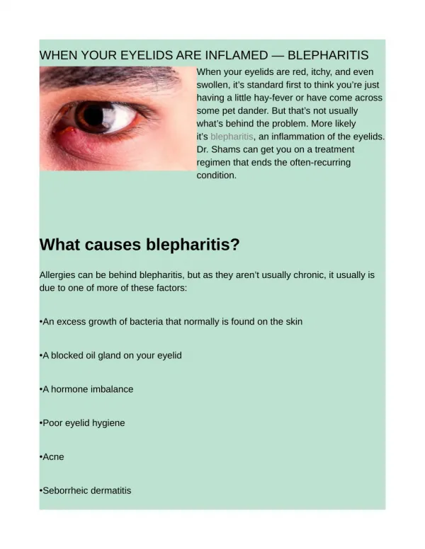
BLEPHARITISBelpharitis usually presents as a chronic blepharoconjunctivis and it is the most common external eye disorder in clinical practice. Types: • Squamous blepharits. • Ulcerative blepharitis. • Squamous Blepharitis : Low grade infection on top of abnormal secretions of the lid glands similar to seborrheic dermatitis.
Clinical picture: Small, white, scales are present between the lashes. Removal of the scales reveals a hyperemic lid margin without ulceration. • Treatment: General treatment of seborrhea. Remove scales with 3% sodium bicarbonate or diluted baby shampoo. Rub antibiotic ointment into the lid margin. The treatment must be prolonged 2 – 3 weeks.
Ulcerative Blepharitis: Etiology: Predisposing Factors: Poor hygiene Malnutrition DiabetiesErrors of refraction Exciting cause : Infection of the lid margin with staphylococcus aureus (sebaceous, sweat gland and hair follicles)
Signs: Yellow crusts glue the lashes together. Minute ulcers of the lid margin which bleed easily when crusts are removed.. Treatment: General treatment :Improve general healthControl diabetes if present.
Local treatment • Lid hygiene. • Frequent massage to evacuate meibomian secretions. • Meticulous removal of scales by scrubbing the lid margins with baby shampoo or 3% sodium bicarbonate lotion. • Elimination of infection
INFLAMMATION OF THE GLANDS OF THE LID Stye: (Hordeolumexternum) Acute suppurative inflammation of Zeis gland and the lash follicle, forming a small abscess.
Etiology: Infection of a Zeiss gland by staphylococcus aureus Clinical picture: Symptoms: Swelling of the lid Severe pain, first dull then throbbing. Signs: Diffuse red swelling Related to a lash Close to the lid margin. Points on the skin side.
Treatment: • Hot fomentations. • Local antibiotic drops and ointment. • Systemic antibiotics. • When pointing occurs, The pus must be evacuated by : • Epilation of the related lash. • Horizontal incision. • For recurrent cases: correct the underlying cause.
2. Hordeoluminternum • Acute suppurative inflammation of the meibomian gland caused by staphylococcus aureus. • It may be primary or it may occur on top of a chronic inflammation of the mebomian gland (chalazion).
Chalazion ( cyst) It is a chronic non-specific inflammatory granuloma of mebomian gland.
Etiology: Etiology is unknown .It is a granuloma produced by the retained contents of the gland following obstruction of its duct, or as a result of chronic irritation by a low virulence organism. Treatment : • Very small chalazion: local antibiotic and steroid preparation. • Marginal chalazion : scraping from lid margin followed by diathermy
Treatment : • Moderate or large chalazion : vertical incision and scraping through the conjunctival side. • Multiple chalazion : combined excision of tarsus and conjunctiva leaving the lower third of the tarsus ( to avoid lid notching) with replacement by a mucous graft from the lip. • Recurrent chalazion of the same gland : excision biopsy to exclude malignant tumor.
DISORDERS OF EYELASHESTrichiasis :Trichiasis is a condition where more than 4 lashes are rubbing against the cornea or conjunctiva. Rubbing lashes: Rubing is a term applied to the condition where 4 lashes or less are misdirected to rub against the cornea or conjunctiva.
DISORDERS OF EYELASHESMadarosis : permanent absence of eye lashes due to destruction of the lash follicles. It may be partial or total.
DISORDERS OF EYELASHESDistichiasis: An extra row of lashes situated in or near to the openings of the meibomian glands.
MALPOSTION OF THE EYELIDENTROPIONDefinition: Entropion is the rolling inwards of the eyelid. The whole row of the lashes will be rubbing against the cornea and finally there will be a deformity of the tarsus.
ENTROPION: Types: • Cicatricial (fibrotic) entropion: Fibrosis of the palpebral conjunctiva due to trachoma, chemical burns, diphtheria and ocular cicatricialpemphigoid. • Spastic entropion: due to spasm of orbicularis muscle in response to ocular irritation e.g inflammation, exposed sutures, operations following enucleation or enophthalmos
Involutional ( senile) entropion: affects only the lower lid due to overriding of the preseptal portion of orbicularis muscle over the pretarsal portion. • Congenital entropion: usually affecting the whole lower eyelid
ECTROPIONDefinition : Ectropion is rolling outwards of the eyelid from the globe. It usually affects the lower lid as it stands against gravity.
ECTROPIONTypes: • Involutional ( senile ) ectropion: due to senile weakness of the orbicularis muscle and relaxation of the palpebral ligaments. • Cicatricial (fibrotic) ectropion: due to scarring and contracture of the skin of the lower lids by burns, trauma or tumor. • Paralytic ectropion: due to paralysis of orbicularis muscle in facial nerve palsy. • Mechanical ectropion:due to increased weight of lower lid by e. g. multiple chalazia. • Congenital ectropion:rare
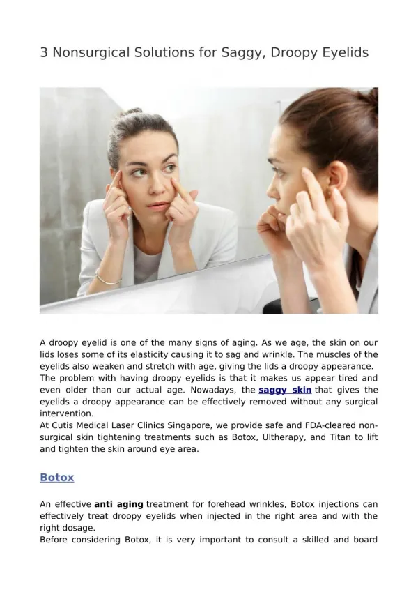
PTOSISDefinition : Ptosis is the drooping of the upper eyelid that normally covers the upper 1/6 of the cornea
Etiology (types) • Myogenic ptosis : due to disorders of levator muscle. • Congenital ptosis: due to dystrophy of levator muscle • Acquired ptosis: myasthenenia gravis: due to a defect at the myoneural junction • Neurogenic (paralytic) ptosis: due to a disorder of nerve supply. • Third nerve palsy: (diabetes, congenital, or traumatic) • Horner's syndrome : due to disorder of sympathetic nerve supply leading to Mullers muscle paralysis. (partial ptosis, miosis, anhydrosis and enophthalmos)
3. Aponeurotic ptosis: due to disorder of levatoraponeurosis. • Involutional (senile) ptosis: degenerative process with age. • Postoperative ptosis: after cataract or retinal detachment surgery . • 4.Mechanical ptosis : Excess weight due to edema, trauma, tumor. Conjunctival scarring.
Xanthelasma: Subcutaneous deposits of cholesterol in the medial canthus region. It is seen in diabetics and in patients with hypercholesterolemia