Download
1 / 92
980 likes | 1.57k Views
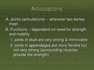
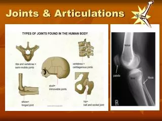
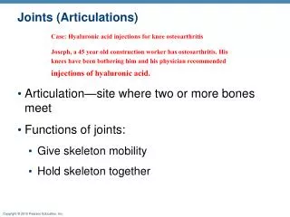
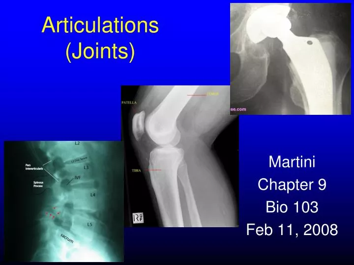
Articulations (Joints). Martini Chapter 9 Bio 103 Feb 11, 2008. Joint Classifications. Functional Classification based on range of motion Anatomical (structural) Classification based on material in joint. Structural vs. Functional Joint Classifications.

E N D
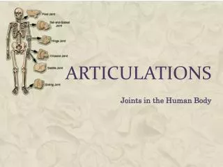
Articulations (Joints) Martini Chapter 9 Bio 103 Feb 11, 2008
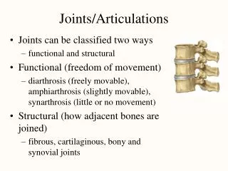
Joint Classifications Functional Classification based on range of motion Anatomical (structural) Classification based on material in joint
Structural vs. Functional Joint Classifications structural categories do have some relationship with the functional categories (structure function)
Structural Joint Categories • Fibrous • no joint cavity • held together with fibrous connective tissue
Structural Joint Categories • Fibrous • no joint cavity • held together with fibrous connective tissue • Cartilaginous • no joint cavity • held together with cartilage
Structural Joint Categories • Fibrous • no joint cavity • held together with fibrous connective tissue • Cartilaginous • no joint cavity • held together with cartilage • Synovial • has a joint cavity • articular capsule and ligaments join bones
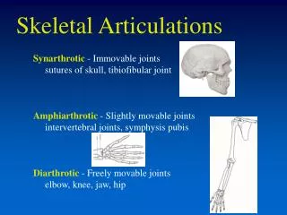
3 Functional Classifications Synarthrosis (together + joint) no movement structural type: fibrous, cartilaginous, or bony fusion
4 Types of Synarthrotic Joints Suture (sewn together) fibrous bound by dense fibrous connective tissue found only in skull
4 Types of Synarthrotic Joints Gomphosis (bolted together) fibrous binds teeth to bony sockets (maxillary bone and mandible) fibrous connection between tooth and socket is called periodontal ligament
4 Types of Synarthrotic Joints Synchondrosis (together + cartilage) cartilaginous rigid bridge between 2 bones epiphyseal cartilage of long bones Between ribs and sternum
4 Types of Synarthrotic Joints Synostosis totally rigid fused bones (bony fusion) epiphyseal lines of mature long bones metopic suture in frontal bone (doesn’t always fuse!)
Craniosynostosis • when skull sutures fuse prematurely
3 Functional Classifications Amphiarthrosis (both sides + joint) little movement structural type: fibrous or cartilaginous
2 Types of Amphiarthroses Syndesmosis (desmos = ligament) bones connected by ligaments example between tibia and fibula
2 Types of Amphiarthroses Symphysis bones separated by a wedge or pad of fibrocartilage intervertebral discs connection between pubic bones
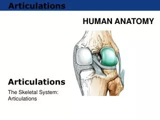
3 Functional Classifications Diarthrosis (through + joint) more movement articular cartilage + synovial fluid + accessory structures
Diarthrosis (Synovial) Joints freely moveable joints catagorized by movement type typically located in appendicular skeleton, at connections of long bones The shoulder joint is the most freely moving joint in the body.
Synovial Joint Structure articular capsule unites 2 bone ends
Synovial Joint Structure articular capsule unites 2 bone ends outer layer fibrous, dense irregular connective tissue that blends with the periostea of the two bones to form ligaments
Synovial Joint Structure articular capsule unites 2 bone ends outer layer fibrous, dense irregular connective tissue that blends with the periostea of the two bones to form ligaments inner layer (synovial membrane) composed of areolar tissue and epithelial tissue
Synovial Joint Structure articular cartilage like hyaline cartilage, but no perichondrium matrix more watery
Synovial Joint Structure synovial fluid thin film between articular cartilage creates smooth gliding surface
Synovial Joint Structure synovial fluid thin film between articular cartilage creates smooth gliding surface similar to interstitial fluid with high concentration of proteoglycans consistency of molasses
Functions of Synovial fluid Lubrication cartilage sucks up fluid like sponge and releases when compressed to reduce friction
Functions of Synovial fluid Lubrication cartilage sucks up fluid like sponge and releases when compressed to reduce friction Nutrient Distribution fluid circulates bringing nutrients and removing wastes for chondrocytes
Functions of Synovial fluid Lubrication cartilage sucks up fluid like sponge and releases when compressed to reduce friction Nutrient Distribution fluid circulates bringing nutrients and removing wastes for chondrocytes Shock absorption lessens shock by distributing pressure evenly across articular surface
Synovial Joint Accessory Structures Cartilages meniscus(crescent) aka articular disc pad of fibrocartilage that sits between 2 bones
Synovial Joint Accessory Structures Fat pads covered by synovial membrane protect articular cartilages act as packing material for joint, filling in spaces left as joint cavity changes shape
Synovial Joint Accessory Structures Ligaments attach bone to bone support and strengthen joints
Synovial Joint Accessory Structures Ligaments Intrinsic (capsular) ligaments thickening of joint capsule
Synovial Joint Accessory Structures Ligaments Extrinsic ligaments separate from joint capsule intracapsular (inside capsule) e.g., ACL, PCL extracapsular (outside capsule) patellar ligament
Sprains • ligament collagen stretched/torn • more likely to break bone than tear ligament
Synovial Joint Accessory Structures Tendons attach to muscles around joint, but not a part of articulation pass through/around joint and can affect joint movement
Synovial Joint Accessory Structures Bursae(pouch) pockets of synovial fluid that cushion areas where tendons or ligaments rub against other tissue synovial tendon sheaths tubular bursae that surround tendons where they cross bone
Bursitis • inflammation of bursae causing pain during motion • caused by: • overuse • pressure • bunion • chemicals/infection
Stabilization of Joints • movement beyond range causes joint damage
Stabilization of Joints • movement beyond range causes joint damage • greater range of motion = greater chance for injury
Stabilization of Joints • movement beyond range causes joint damage • greater range of motion = greater chance for injury • To reduce chance of injury joints are stabilized by:
Stabilization of Joints • movement beyond range causes joint damage • greater range of motion = greater chance for injury • To reduce chance of injury joints are stabilized by: • collagen fibers of capsule and ligaments • shape of articulating surfaces and menisci might prevent movement in certain directions • presence of other bones, muscles, fat pads around joint • tension in tendons encourages movement in specific direction
Describing Dynamic Motion 3 possible movements 1. linear (gliding) 2. angular (circumduction) 3. rotation
Types of Movements at Synovial Joints Linear Motion (Gliding) 2 opposing surfaces slide past one another between carpal bones clavicle and sternum between tarsal bones
Types of Movements at Synovial Joints Flexion/Extension movement in anterior/posterior plane flexion reduces the angle extension increases the angle in the anatomical position, all major joints (except ankle) are at full extension. extension past the anatomical position is called hyperextension example is bending your neck backwards to look at the sky
Types of Movements at Synovial Joints Abduction/Adduction angular movement in frontal plane abduction moves away from the longitudinal axis of the body adduction moving towards the longitudinal axis of the body
Types of Movements at Synovial Joints Circumduction angular motion, without rotation
Types of Movements at Synovial Joints Rotation Left or right rotation Medial rotation (inward rotation) rotates toward axis Lateral rotation (outward rotation) rotates away from axis
Types of Movements at Synovial Joints Rotation of the forearm Pronation: rotates forearm, radius over ulna Supination: forearm in anatomical position
Special Movements Inversion/Eversion twisting motion of the foot that turns sole inward (inversion) or outward (eversion)
Special Movements Dorsiflexion/Plantar flexion flexion at ankle joint dorsiflexion elevates sole plantar flexion elevates heel NOTE: it is acceptable to use the terms flexion and extension for these movements of the ankle.