Download
1 / 3
30 likes | 140 Views
Supplementary Fig. 1. GRIM-19 G139V. GRIM-19 Y143D. GRIM-19 G139R. GRIM19 Y143A. GRIM-19. Blot : myc. GRIM-19. BN-PAGE. com II. SDS-PAGE. myc.

E N D
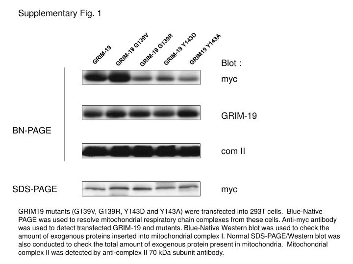
Supplementary Fig. 1 GRIM-19 G139V GRIM-19 Y143D GRIM-19 G139R GRIM19 Y143A GRIM-19 Blot : myc GRIM-19 BN-PAGE com II SDS-PAGE myc GRIM19 mutants (G139V, G139R, Y143D and Y143A) were transfected into 293T cells. Blue-Native PAGE was used to resolve mitochondrial respiratory chain complexes from these cells. Anti-myc antibody was used to detect transfected GRIM-19 and mutants. Blue-Native Western blot was used to check the amount of exogenous proteins inserted into mitochondrial complex I. Normal SDS-PAGE/Western blot was also conducted to check the total amount of exogenous protein present in mitochondria. Mitochondrial complex II was detected by anti-complex II 70 kDa subunit antibody.
Supplementary Fig. 2 NDUFS3 1-60-HA GRIM-19-HA NDUFA9 1-100-HA NDUFA9 1-80-HA NDUFA9 1-60-HA NDUFS3 1-100-HA NDUFS3 1-80-HA Blot : HA BN PAGE GRIM-19 comII 70 kDa subunit HA SDS PAGE NDUFA9 mutants (1-100 and 1-80) and NDUFS3 mutant (1-100) can be assembled into mitochondrial complex I. WT-GRIM-19, NDUFA9 mutants (1-100, 1-80 and 1-60), and NDUFS3 mutants (1-100, 1-80 and 1-60) were transfected into 293T cells. The same procedure was conducted as in Supplementary Fig 1.
Supplementary Fig. 3 IPHA Complex I Mitochondrial complex I was immunoprecipated by complex I capture kit (MitoSciences (Eugene, OR, USA) according to manufacturer’s instructions. DN-GRIM-19-HA incorporated into complex I was immunoprecipated by HA-conjugated agarose beads. The immunoprecipated protein was eluted from the beads and subjected to SDS-PAGE. The gel was visualized by silver stain. DN-GRIM-19