Download
1 / 33

E N D
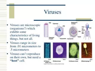
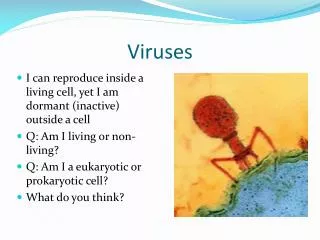
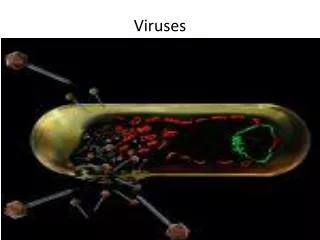
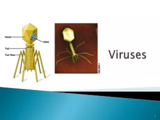
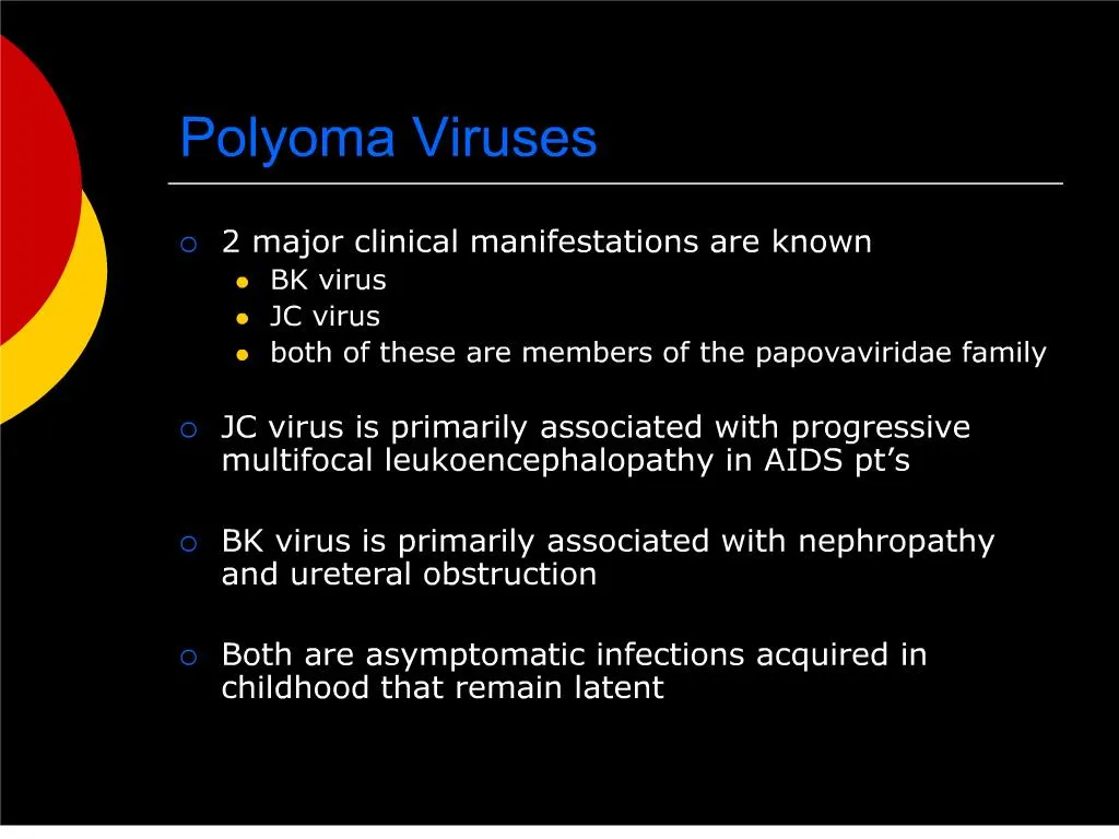
1. Polyoma Viruses 2 major clinical manifestations are known
BK virus
JC virus
both of these are members of the papovaviridae family
JC virus is primarily associated with progressive multifocal leukoencephalopathy in AIDS pt�s
BK virus is primarily associated with nephropathy and ureteral obstruction
Both are asymptomatic infections acquired in childhood that remain latent
3. BK virus Viruria can be detected in many populations, but the most clinically important is renal transplant recipients
Exact pathogenesis of the infection is poorly understood
Diagnosis must be made histologically (biopsy)
Special stains and PCR can help solidify the diagnosis
4. BK virus Treatment
Only effective therapy is immune reconstitution (i.e. reduction of immunnosuppressant therapy)
Cedofivir and leflunomide are effective in reducing viral load, but do very little to change the course of disease, and are both nephrotoxic
One must effectively walk the tightrope between progressive renal destruction secondary to infection, and acute rejection causing graft loss
5. BK virus Several case reports exist for BK nephropathy in non-renal transplant recipients
Bone marrow
Heart
Lung
However, these are rare, and are at level of case report
6. Learning Objectives Recognize the 4 categories of infections in transplant recipients
Segregate and stratify patient�s infection risk profile by the time from transplantation
Be focally empiric and aggressive to make a definitive diagnosis in transplant patients
7. Intro Recognizing the presentation of infections in transplanted patients is paramount
However, immunosuppressed patients often present atypically, and with far advanced stages of infection
Fever is a non-specific finding, which can be attributed to medications and acute rejection
Invasive testing is often required to make an accurate and timely diagnosis
8. Epidemiology The risk, rate, and type of infection vary over time from transplantation
Currently, there are no assays to measure risk/susceptibility to infection
Risk of infection is an interplay between
Exposure history (of donor and recipient)
Intensity and quality of immunosuppression
Use of prophylactic medications
9. Classification of Infections Donor-derived
Recipient-derived
Nosocomial
Community-acquired
10. Donor-derived Infection Most are latent
CMV, TB, T.cruzi
Rarely can be acute
Bacteremia/viremia at time of procurement
West Nile, rabies, HIV, hepatitis, LCV
The majority of these are sub-clinical in healthy patients, but can be catastrophic when transplanted into an immunosuppressed patient
At present, routine evaluations of donors for infectious diseases relies upon serologic antibody testing, and thus sensitivity is not 100% for those that may not have had time to seroconvert
11. Donor-derived Transplantation of organs from deceased donors with viral syndromes is controversial
Livers with known Chagas or Hep B infection may be used as there are effective treatments for these infections
Hep C infected organs are sometimes transplanted into Hep C(+) donors
13. Recipient-derived Infections Infections that can be treated or controlled do not necessarily preclude transplantation
Most commonly screened for:
TB
syphilis
Viral: CMV, EBV, VZV, HSV, HIV, HBV, HCV
Other things to think of
T.cruzi, strongyloides, cryptococcus
Endemic fungi: histoplasma, coccidioides, paracoccidioides, aspergillus, blastomycosis
15. Immunizations Pt�s should be current on the following vaccines
MMR
HBV
Influenza
Strep pneumoniae
Tetanus
Diphtheria
Pertussis
Polio
VZV � if never infected
Consideration should be given to boosters for any of the above prior to transplantation as live vaccines are generally contraindicated post-transplant, and immunologic memory will become impaired
16. Peri-operative Prophylaxis Liver
Skin, enterococcus, anaerobes, enterobacteriaceae
Most common site of invasive fungal infxn
Lung
GNR, molds, endemic fungi
MRSA and VRE according to antiobiogram prevalence
Second most common site of invasive fungal infxn
17. Nosocomial Infections MRSA
VRE
fluconazole-resistant Candida species
associated with surgical site and indwelling catheters
C.diff
Resistant gram-negative bacilli
Aspergillus
18. Community Infections Aspergillus
Nocardia
Cryptococcus neoformans (birds)
Respiratory viruses
Secondary bacterial superinfection
19. Monitoring Immunosuppression There are no specific tests currently available to determine the overall susceptibility of patients to infection�
�but they are on the horizon
Currently, the known determinants contributing to the overall risk of infection are the dose, duration, and sequence of immunosuppressive therapies
20. Figure 3. Dynamic Assessment of the Risk of Infection after Transplantation. The risk of infection is a function of the net state of immunodeficiency. The presence of specific, common infections can be detected by means of quantitative assays measuring nucleic acids or proteins derived from potential pathogens. Multiple simultaneous quantitative (multiplex) assays can be performed diagnostically in a single sample with the use of polymerase chain reaction. Each line represents a single patient's sample (Panel A). The presence of specific infections can be assessed with the use of genomic arrays measuring the up-regulation or down-regulation of host genes during infection (Panel B, courtesy of Shaf Keshavjee, M.D., University of Toronto). Lytic and latent epitopes are viral antigens presented in either the lytic or latent phase of Epstein-Barr virus (EBV) infection. The transplant recipient's cellular immune response to specific pathogens such as EBV can be determined by measurements of cellular activation by pathogen-specific antigens (Panel C, courtesy of Christian Brander, Massachusetts General Hospital). The factors contributing to the degree of immunologic impairment and standard assays that assess the patient's risk of infection will be supplemented in the future by new quantitative measures of allograft- and pathogen-specific immune function and the risk of infection (Panel D). RFU denotes relative fluorescence units, CMV cytomegalovirus, BK polyomavirus type BK, HHV-6 human herpesvirus 6, HHV-7 human herpesvirus 7, PBMCs peripheral-blood mononuclear cells, SLE systemic lupus erythematosus, HCV hepatitis C virus, and HBV hepatitis B virus.Figure 3. Dynamic Assessment of the Risk of Infection after Transplantation. The risk of infection is a function of the net state of immunodeficiency. The presence of specific, common infections can be detected by means of quantitative assays measuring nucleic acids or proteins derived from potential pathogens. Multiple simultaneous quantitative (multiplex) assays can be performed diagnostically in a single sample with the use of polymerase chain reaction. Each line represents a single patient's sample (Panel A). The presence of specific infections can be assessed with the use of genomic arrays measuring the up-regulation or down-regulation of host genes during infection (Panel B, courtesy of Shaf Keshavjee, M.D., University of Toronto). Lytic and latent epitopes are viral antigens presented in either the lytic or latent phase of Epstein-Barr virus (EBV) infection. The transplant recipient's cellular immune response to specific pathogens such as EBV can be determined by measurements of cellular activation by pathogen-specific antigens (Panel C, courtesy of Christian Brander, Massachusetts General Hospital). The factors contributing to the degree of immunologic impairment and standard assays that assess the patient's risk of infection will be supplemented in the future by new quantitative measures of allograft- and pathogen-specific immune function and the risk of infection (Panel D). RFU denotes relative fluorescence units, CMV cytomegalovirus, BK polyomavirus type BK, HHV-6 human herpesvirus 6, HHV-7 human herpesvirus 7, PBMCs peripheral-blood mononuclear cells, SLE systemic lupus erythematosus, HCV hepatitis C virus, and HBV hepatitis B virus.
21. Figure 4. Changing Timeline of Infection after Organ Transplantation. Infections occur in a generally predictable pattern after solid-organ transplantation. The development of infection is delayed by prophylaxis and accelerated by intensified immunosuppression, drug toxic effects that may cause leukopenia, or immunomodulatory viral infections such as infection with cytomegalovirus (CMV), hepatitis C virus (HCV), or Epstein-Barr virus (EBV). At the time of transplantation, a patient's short-term and long-term risk of infection can be stratified according to donor and recipient screening, the technical outcome of surgery, and the intensity of immunosuppression required to prevent graft rejection. Subsequently, an ongoing assessment of the risk of infection is used to adjust both prophylaxis and immunosuppressive therapy. MRSA denotes methicillin-resistant Staphylococcus aureus, VRE vancomycin-resistant Enterococcus faecalis, HSV herpes simplex virus, LCMV lymphocytic choriomeningitis virus, HIV human immunodeficiency virus, PCP Pneumocystis carinii pneumonia, HBV hepatitis B virus, VZV varicella-zoster virus, SARS severe acute respiratory syndrome, PML progressive multifocal leukoencephalopathy, and PTLD post-transplantation lymphoproliferative disorder. Modified from Fishman and Rubin1 and Rubin et al.45Figure 4. Changing Timeline of Infection after Organ Transplantation. Infections occur in a generally predictable pattern after solid-organ transplantation. The development of infection is delayed by prophylaxis and accelerated by intensified immunosuppression, drug toxic effects that may cause leukopenia, or immunomodulatory viral infections such as infection with cytomegalovirus (CMV), hepatitis C virus (HCV), or Epstein-Barr virus (EBV). At the time of transplantation, a patient's short-term and long-term risk of infection can be stratified according to donor and recipient screening, the technical outcome of surgery, and the intensity of immunosuppression required to prevent graft rejection. Subsequently, an ongoing assessment of the risk of infection is used to adjust both prophylaxis and immunosuppressive therapy. MRSA denotes methicillin-resistant Staphylococcus aureus, VRE vancomycin-resistant Enterococcus faecalis, HSV herpes simplex virus, LCMV lymphocytic choriomeningitis virus, HIV human immunodeficiency virus, PCP Pneumocystis carinii pneumonia, HBV hepatitis B virus, VZV varicella-zoster virus, SARS severe acute respiratory syndrome, PML progressive multifocal leukoencephalopathy, and PTLD post-transplantation lymphoproliferative disorder. Modified from Fishman and Rubin1 and Rubin et al.45
22. Early post-transplant period (30d) Opportunistic infections are rare in the first month post-transplant
>1 month of medical therapy is required to effectively deplete cell-mediated therapy
One exception is large, prolonged doses of corticosteroids
Infections are generally donor-derived or associated with complications from the surgery itself
24. Intermediate period (1-6mos) Viral infections and allograft rejection account for the majority of febrile episodes
Adherence to Bactrim and antiviral prophylaxis renders infections such as PCP, UTI�s, listeria, toxoplasmosis, and herpes very unlikely
Fungi, cryptococcus, T.cruzi, strongyloides can surface
Other: polyoma virus (BK and JC), recurrent HCV
26. Late post-transplant period (>6mos) Risk wanes as immunosuppressive therapy is tapered
Risk profile however, is �reset� with each episode of acute rejection
Chronic viral infections can cause allograft injury
HCV ? cirrhosis
BOOP in lungs
CMV ? coronary vasculopathy
PTLD
Skin/anogenital cancers
Fungi/molds, virusesn and �typical� bugs still remain on radar
28. Long-term Prophylaxis Bactrim for at least 3 months, sometimes for life
CMV and HSV prophylaxis is not standardized, and varies according to immunnosuppressive regimen and institution
Rarely chronic suppressive antifungal Rx for pt�s with history of significant disease
29. General Lifestyle Rec�s Avoid sick contacts, esp respiratory
Avoid dusty sites (i.e. construction sites)
Avoid ingestion of well and lake water
Avoid undercooked meats
Avoid soft cheeses and unpasteurized dairy products
Avoid unwashed fruits/veggies
30. Back to our patient Due to the multifactorial nature of his renal failure, there is little hope for regaining renal function
He is currently using peritoneal dialysis and considering being listed for renal transplant
31. Summary Be aware of the time frame from transplant leading you to the most likely type of infection
<30d think nosocomial or donor-derived
1-6mos, reactivation of viruses and atypical infections �classic� for transplant pt�s
>6mos, think of �typical� or community-acquired bugs as their risk returns to somewhat normal
32. References Fishman JA. NEJM 2007;357:2601-14
http://content.nejm.org/cgi/content/full/357/25/2601
33. Figure 2. Assessment of the Risk of Infection at the Time of Transplantation. The risk of infection transmitted from the organ donor or activated in the recipient can be assessed at the time of transplantation. Donor and recipient screening are based on the epidemiologic history and serologic testing. The use of sensitive molecular and protein-based assays may enhance the safety of organ transplantation while expanding the use of potentially infected grafts. The transplant recipient's risk is a function of the technical outcome, epidemiologic factors, and the intensity of immunosuppression. VDRL denotes Venereal Disease Research Laboratory test, HIV human immunodeficiency virus, CMV cytomegalovirus, EBV Epstein-Barr virus, HSV herpes simplex virus, VZV varicella-zoster virus, HBV hepatitis B virus, HBsAg hepatitis B surface antigen, anti-HBsAg antibodies against hepatitis B surface antigen, and HCV hepatitis C virus.Figure 2. Assessment of the Risk of Infection at the Time of Transplantation. The risk of infection transmitted from the organ donor or activated in the recipient can be assessed at the time of transplantation. Donor and recipient screening are based on the epidemiologic history and serologic testing. The use of sensitive molecular and protein-based assays may enhance the safety of organ transplantation while expanding the use of potentially infected grafts. The transplant recipient's risk is a function of the technical outcome, epidemiologic factors, and the intensity of immunosuppression. VDRL denotes Venereal Disease Research Laboratory test, HIV human immunodeficiency virus, CMV cytomegalovirus, EBV Epstein-Barr virus, HSV herpes simplex virus, VZV varicella-zoster virus, HBV hepatitis B virus, HBsAg hepatitis B surface antigen, anti-HBsAg antibodies against hepatitis B surface antigen, and HCV hepatitis C virus.