Download
1 / 25
510 likes | 3.86k Views
Fig 23.1. Digestive System. Digestive System. Organs of the digestive system GI Tract Organs - mouth, pharynx, esophagus, stomach, small intestine, large intestine GI tract also called the alimentary canal, digestive tract or gut

E N D
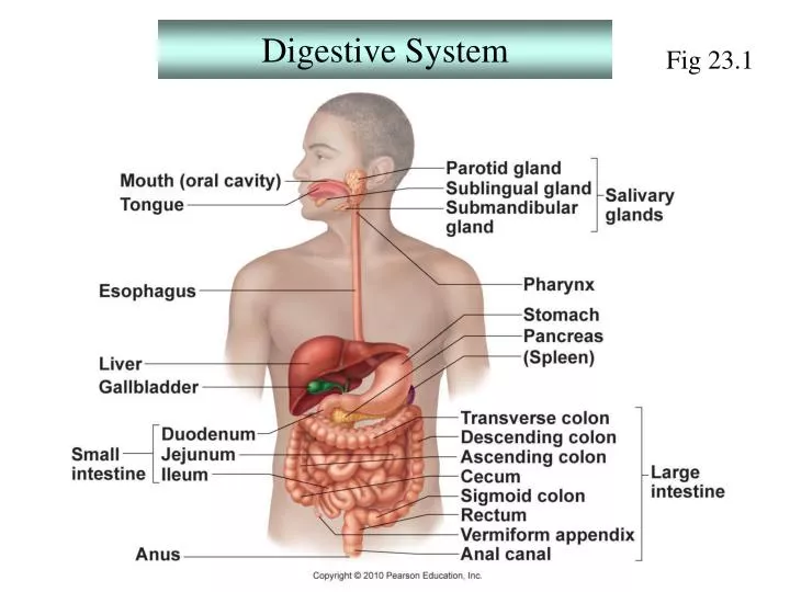
Fig 23.1 Digestive System
Digestive System Organs of the digestive system • GI Tract Organs-mouth, pharynx, esophagus, stomach, small intestine, large intestine GI tract also called the alimentary canal,digestive tract or gut • Accessory Digestive Organs- teeth, tongue, salivary glands, gall bladder, liver, pancreas
Gastrointestinal Mucosa- moist covering and liningmembrane lining the inside of the GI tract absorbs nutrients- aided by large surface area due to villi and microvilli esp. in sm. intestine secretes mucin —> mucous for lubrication, protection protects- mucosal immune functions (MALT) gastric mucosahas specialized secretory cells
Digestive System Gastrointestinal Tract Activities/Processes 1.Ingestion-intake of food 2.Propulsion-movement of food through the GI tract Peristalsis-alternative contraction and relaxation of adjacent sections of the alimentary tract resulting in food propulsion 3. Mechanical digestion-chewing,mixing, stomach churning, segmentation Segmentation-alternative contraction and relaxation of non- adjacent sections of the alimentary tract, resulting in mixing of food 4. Chemical digestion-catabolic steps which break down complex molecules to monomers or fragments which can be absorbed by the GI tract 5. Absorption uptake of nutrients from the lumen of the GI tract into blood or lymph via passive and active transport 6. Defecation- elimination of indigestible substances from the body
Overview of the functions of the Gastrointestinal Tract Table 23.2
Microscopic Anatomy of the stomach *read pp 869 to 871up to “Digestive Processes….” Secretory cells of the gastric pits: Know what Chief cells, Parietal cells and Enteroendocrine cells of the stomach mucosa are and their function(s)
The small intestineduodenum, jejunum, ileum Digestion is completed and most absorption occurs here Villi-fingerlike projections containing underlying: Capillary bed- Absorbs most nutrients and water soluble drugs and some lipophilic drugs Lymphatic capillary (lacteal)- Absorbs fats and highly lipophilic drugs
Accessory Digestive Organs: Liver, Gall Bladder, Pancreas 1. Liver-largest gland in the body located below the diaphragm-has numerous functions function in GI tract processes-production of bile, a yellow-green alkaline fluid containing bile salts that is released into the small intestine (duodenum) via the bile duct; main function of bile →is to emulsify fats
Liver Functions • carbohydrate metabolism/storage • detoxification • serum protein production • hormone inactivation • protein metabolism/urea production • lipid metabolism • iron recycling • cholesterol synthesis
Structure of the liver- A. gross anatomy 4 lobes-left, right,caudate and quadrate functional units- liver lobules B. microscopic anatomy of liver lobules-hepatocytes- mature liver cells that perform most of the liver functions arranged in ‘plates’; high regeneration capacity
B.microscopic anatomy of liver lobules optionalFig 23.25 hepatocytes- Perform main Functions of the Liver.
B.microscopic anatomy of liver lobules hepatocytes- mature liver cells that perform most of the liver functions arranged in ‘plates’; high regeneration capacity
Accessory Digestive Organs: Liver, Gall Bladder, Pancreas 2. Gall Bladder-thin-walled muscular sac located below the ventral portion of the liver that stores and concentrates bile bile transferred from liver to gall bladder viacommon hepatic duct 3. Pancreas- digestive function-produces pancreatic juice containing digestive enzymes (proenzymes) for release into duodenum via the main and accessory ducts
Flowchart of chemical digestion and absorption of foodstuffs optional Fig. 23.32