Download
1 / 63
640 likes | 898 Views
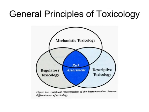
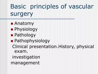
Principles of Vascular Toxicology. Mitchell Troutman, D.V.M. The University of North Carolina at Chapel Hill. Basic Vascular Structure:. Endothelium Smooth Muscle Connective Tissue. Basic Vascular Function. Arteries, veins, and capillaries Conduct blood to tissues Transport oxygen

E N D
Principles of Vascular Toxicology Mitchell Troutman, D.V.M. The University of North Carolina at Chapel Hill
Basic Vascular Structure: Endothelium Smooth Muscle Connective Tissue
Basic Vascular Function • Arteries, veins, and capillaries • Conduct blood to tissues • Transport • oxygen • nutrients • wastes • drugs/chemicals (endogenous and exogenous) • Lymphatics • Return interstitial tissue fluid to blood
Endothelial Cell Properties & Functions • Maintenance of permeability barrier • Anticoagulant/antithrombotic molecules • prostacylcin, thrombomodulin, plasminogen activator (PA), heparin-like molecules • Prothrombotic molecules • von Willebrand factor, tissue factor, PA inhibitor • Extracellular matrix production • collagen, proteoglycans
Endothelial Cell Properties & Functions • Modulation of blood flow & vascular reactivity • vasoconstrictors (endothelin, ACE {Angiotensin converting enzyme}) • vasodilators (nitric oxide, prostacyclin) • Regulation of inflammation & immunity • adhesion molecules, interleukins, histocompatibility antigens • Regulation of cell growth • growth stimulators (PDGF, CSF, FGF) • growth inhibitors (heparin, TGF-) • Oxidation of low-density lipoprotein (LDL)
Vascular Smooth Muscle Cells • Vasoconstriction/vasodilation • in response to normal or pharmacologic stimuli • Synthesis of connective tissue • collagen, elastin, proteoglycans • Release of growth factors & cytokines • Migration & proliferation
Manifestations of Vascular Dysfunction • Structural • arteriosclerosis/atherosclerosis • vasculitis • prothrombotic events • Functional (physiologic) • hypertensive arteriosclerosis • prothrombotic events
Normal Coronary Artery Atherosclerosis Coronary Artery with plaque formation and narrowing of vascular lumen
Pathogenesis of Atherosclerosis • Response to injury hypothesis • injury to arterial endothelium (inc. hypertension) • increased permeability to plasma constituents • adherence of blood monocytes (+ migration) and platelets • lipid accumulation in macrophages foam cells • migration and proliferation of smooth muscle cells deposition of collagen and proteoglycans • repeated injury atheromatous plaque forms
Atherosclerosis: Importance in Toxicology • Toxicants can accelerate or exacerbate • cadmium, selenium, chronic copper toxicity • carbon monoxide • cholesterol and oxygenated derivatives • homocysteine • Major therapeutic area of interest
Hypertension (high Blood Pressure) • Influenced by many factors • cardiac output, vascular compliance, renal disease • Blood vessels become thickened and less distensible heart works harder to circulate blood • Chronic damage and cardiovascular failure can occur
Hypertensive Arteriosclerosis • Pathogenesis: • inadequate renal sodium excretion increased neurohormonal release • increased plasma and ECF volume • increased cardiac output HYPERTENSION that leads to chronic vascular endothelial damage increased natriuretic hormone increased total peripheral resistance
Hypertensive Arteriosclerosis Canine glomerular hyaline arteriosclerosis, PAS stain, 200X
Hypertensive Arteriosclerosis Normal Coronary Artery Coronary Artery branch with hyperplastic arteriosclerosis and near occlusion of vascular lumen
Hypertensive Arteriosclerosis: Importance in Toxicology • Toxicants can accelerate or exacerbate • cadmium, arsenic, mercury • allylamine, chlorophenoxy herbicides • organophosphates • Can accelerate atherosclerosis and potentiate cerebrovascular hemorrhage
What is drug-induced vasculitis? • Vasculitis: inflammation of blood vessels • Vasculitis can occur either: • Spontaneously – Canine polyarteritis nodosa (i.e., canine pain syndrome) • After exposure to exogenous agents - Infection agents - Pharmaceuticals (i.e., drug-induced)
Drug-induced vasculitis: In Humans • Most drug-induced vasculitides are non-necrotizing hypersensitivity type (e.g., binding of drug/hapten + protein/carrier antigen), involving small and medium arteries and veins and spare large arteries. Localized mainly to the skin (maculopapular rashes followed by palpable purpura), often associated with systemic symptoms (e.g., arthralgia, malaise, fever); end-stage organ damage (e.g., lungs, kidneys) possible from immune-complex deposition Examples: • Many drugs ( e.g., antibiotics, propylthiouracil, hydralazine) • Biologics (e.g., hematopoietic growth factors, interferons, mAbs) ANIMALS ARE NOT GOOD PREDICTORS OF IMMUNE-MEDIATED VASCULITIS IN HUMANS
Drug-induced vasculitis: In animals • Most common is: Due to drug- induced hemodynamic alterations (vasoactive arteriopathy). Usually involves medium to large sized vessels, resulting from functional damage associated with excessive hemodynamic activity • Less common are: • Due to exacerbation of spontaneous disease • Due to primary cytotoxicity (toxic vasculitis, like allylamine and acrolein) • Due to immune-mediated mechanism (hypersensitivity vasculitis), affecting thin-walled vessels, sparing muscular arteries End-stage organ damage is rare
Drug-induced hemodynamic alterations (vasoactive arteriopathy) Pharmacological agents inducing vasoactive arteriopathy are divided into: • Vasodilators and positive inotropic agents – Pathology: Rat – Splancnic, renal and ovarian large muscular arteries Dog - coronary vessels Examples are as follow: Dopaminergics agonist – Fenoldopam, dopamine Dopamine ß-hydroxylase inhibitors – SKF 102698 Phosphodiesterase (PDE) inhibitors - theophylline Serotoninergic compounds – SKF 103829 Endothelin A receptor antagonists – bosentan Possible mechanisms: Prolonged vasodilatation associated with increased regional blood flow and vessel wall stress • Vasoconstrictors – Endothelin-1, noradrenaline and digoxin (coronary arteritis in dog) Monocrotaline (pulmonary arteritis in rats)
Theophylline (a nonspecific PDE inhibitor) induce splanchnic arteriopathy in rats Nyska A, Herbert RA, Chan PC, Haseman JH, Hailey JR (1998) Theophylline-induced mesenteric periarteritis in F344/N rats. Arch. Tox. 72:731-737
Theophylline (1,3-dimethyxanthine) • An alkaloid found in cocoa and tea • Structurally related to caffeine and theobromine • Used as bronchodilator such as in asthma and myocardial stimulation • In the NTP studies - The compound was administered by gavage to B6C3F1 mice and F344 rats, but vascular lesions were seen only in rats (mainly males) in the 16-D, 14-W and 2-Y, at doses of 75 mg/kg • Vascular lesions affected the medium and large splanchnic arteries
Hemorrhage and necrosis within the media of a mesenteric artery of male rat given 400 mg theophylline/kg BW for 16 D
N H N H Hemorrhage (H) and necrosis (N) within the media of a mesenteric artery of male rat given 400 mg theophylline/kg BW for 16 D Note the damage is located at arterial bifurcation (mesenteric artery and 1st branch)
Periarteritis in pancreatic arteries of male F344 rat given 75 mg/kg of theophylline for two years. Thickened wall Thickened media (hypertrophic smooth muscle cell and fibrosis) Thickened adventitia (fibrosis)
vasoactive arteriopathy Mechanistic studyPhosphodiesterase III inhibitor-induced mesenteric arteriopathy in rats Ref: Joseph EC, Rees JA, Dayan AD. (1996). Mesenteric arteriopathy in the rat induced by phosphodiesterase III inhibitors: an investigation of morphological, ultrastructural, and hemodynamic changes.Toxicol Pathol. 1996 Jul-Aug;24(4):436-450.
SK&F 95654 (PDE III inhibitor)-induced arteriopathy model in rat – pathogenetic investigation • Used as potent inotropic/vasodilator • Pharmacologically: PDE III inhibition increases cAMP in the arterial smooth muscle PATHOLOGY: • Dogs: Epicardial arteriopathy • Rats: Focal or segmental medial necrosis or hemorrhage in splanchnic arteries (100-800 micron in diameter) • Objective: Follow the development of time-course (up to 24 hours) changes in peripheral systolic blood pressure and splanchnic arterial lesions
control high dose Drug-induced decrease in systolic blood pressure over 24h following subcutaneous administration of Vehicle control (, DMSO) and SK&F 95654 at 0.174mmol/kg () and 0.697mmol/kg ().
Time-course development of histopathological and SEM findings • 1st changes after 6 h – endothelial raising and pronounced interendothelial projections • 12 h postdosing – medial hemorrhage and medial compression, degeneration and necrosis • 16 h postdosing – endothelial necrosis, adhesion of leukocytes and activated platelets • 24 h postdosing – medial necrosis and infiltration of inflammatory cells with RBCs
Large intestine first branches of mesenteric artery Small intestine Mesenteric arcade from a rat showing severe grade lesion. Multiple large foci of Hemorrhage are present on all first-branch arteries. Note absence of hemorrhage in superior mesenteric artery (sm) but presence of lesion at junction with first-branch
first branch of mesenteric artery Macroscopic photograph of first-branch mesenteric artery from a rat showing Severe Grade hemorrhagic lesion
SEM, normal endothelial surface of first-branch mesenteric artery from an untreated rat showing confluent layer of interdigitating endothelium (E) with clearly defined plasmalemma ridges.
SEM, focal interendothelial gaps formation - endothelial surface of first-branch mesenteric artery from rat 6 hr following sc does of 0.697 mmol/kg SK&F 95654 showing raised endothelium (E) and multiple interendothelial projections
Pathogenesis and comparative aspects The study suggests that: • Interendothelial gaps are consequence of passive stretching of the endothelium as a result of vasodilatation and associated increased intramural tangential stress • The damage occurs when the critical intramural tension has been exceeded • There was a close correlation between the magnitude and duration of hypotension and severity of arterial lesions
Joseph EC (2000). Arterial lesions induced by phosphodiesterase III (PDE III) inhibitors and DA(1) agonists. Toxicol Lett. 15;112-113:537-46
What does drug-induced vasculitis in animal studies mean to patients(extracted from ”Drug-induced vasculitis: FDA’s perspectives”, presented by Thomas Papoian, Senior pharmacologist FDA, at Toxicology Forum, Aspen, July, 2004 (http://www.toxforum.com) • Possibly nothing, if vascular effects in animals do not occur in patients under therapeutic conditions or exposure - Theophylline showed vasculitis in rats, but has been prescribed in human for many years without any apparent vascular toxicity - (assuming that post-marketing surveillance mechanisms capable of detecting a drug-related signal in people with cardiovascular disease)
What does it mean to patients (extracted from ”Drug-induced vasculitis: FDA’s perspectives”, presented by Thomas Papoian, Senior pharmacologist FDA, at Toxicology Forum, Aspen, July, 2004 (http://www.toxforum.com )– Cont. • But if these specific drug-induced vascular effects can or do occur in patients, then there may be a significant cause for concern because: • Vascular inflammation predisposes to: = progression of atherosclerosis (considered an inflammatory disease) = Rupture of vulnerable plaques = Increased incidence of cardiovascular events: heart attack, stroke, or death • No good way to monitor for vascular inflammation in patients (i.e., no validated specific biomarkers) THE QUESTION: Can certain drugs found to produce vasculitis in animals contribute to the arteriosclerotic process in human?
Approved drugs that cause vascular (vasoactive) injury • Minoxidil • Adenosine • Hydralazine • Milirone • Cilomilast (PDE IV) • Fenoldopam • Bosentan • Theophylline • Caffeine • Nocorandil
Particle matter air pollution size distribution Particles are classified according to their median aerodynamic diameter: Thoracic particles- PM10-deposit in the upper tracheobronchial tree Coarse fraction – PM 10 to 2.5 (10 to 2.5 μM - nanometer). Predominantly natural sources – as soil and grinding Fine particles - PM 2.5 (<2.5 μM). Originate from combustion sources, include primary and secondary particles Ultrafine particles – PM 0.1 (<0.1 μM). Originate from combustion sources, deposits in the alveoli, able to pass directly to the circulatory system from the alveoli
The triggering effect of air pollution in coronary atherosclerosis and acute myocardial infarction • Atherosclerosis is inflammatory – degenerative disease of the arteries • Initiating atherosclerotic factors are for example, disturbed coronary blood flow and low shear stress • The air pollution may trigger the atherosclerosis process, and cause the rupture of quiescent focal atherosclerotic lesion (vulnerable plaque disruption) by inducing or exacerbating one or more of the following effects: • Pro-thrombosis effect (e.g., increased fibrinogen, increased viscosity of the plasma, platelet activation) • Vasoconstrictive effect due to release of the vasoconstrictors (endothelins) • Pro-inflammatory effect (e.g., local or systemic inflammation via action of cytokines, chemokines and Reactive Oxygen Species)
Extracted from: Review : Potential Role of Ultrafine Particles in Associations between Airborne Particle Mass and Cardiovascular Health. Ralph J. Delfino, Constantinos Sioutas, and Shaista Malik. EHP, August 2005
Introduction Epidemiological studies demonstrated an association between exposure to high levels of ambient particulate matter (PM) and increased morbidity and mortality of cardiopulmonary disease in human
Findings in NTP studies with particulatesinhalation exposure for 2 years of B6C3F1 mice • Highly significant association between exposure to indium phosphide and cobalt sulfate heptahydrate (particulate size 1.1-1.8 micron) and incidences of coronary and renal arteritis • Marginal significant association between exposure vanadium pentoxide and gallium arsenide (particulate size 1.1-1.8 micron) and incidences of coronary and renal arteritis
Aspects of indium phosphide- induced arteritis in mice - mononuclear cell arteritis, fibrinoid necrosis, smooth muscle proliferation
Suggested mechanisms • Hypoxia (severe lung space occupying pathology) • Autonomic nervous system activation leading to changes in heart rate (conductive arrythmia and depression in heart rate) or modulation of the coronary vascular tone inducing vasodilation as in the case of PDE III inhibitors • Changes in plasma viscosity (increased plasma fibrinogen) • Increased cytokine expression in the lungs (IL-6)