Download
1 / 26
260 likes | 552 Views
Gene Expression Signatures as Bioindicators of Exposure and Health Effects Related to Environmental Contaminants. Ecosystem & Human Health Risk Assessment. Risk Assessor / Risk Manager Dialogue. Identify Goals and Assessment Endpoints Preparing Conceptual Model Developing Analysis. Analysis.

E N D
Gene Expression Signatures as Bioindicators of Exposure and Health Effects Related to Environmental Contaminants
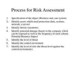
Ecosystem & Human Health Risk Assessment • Risk Assessor / Risk Manager Dialogue • Identify Goals and Assessment Endpoints • Preparing Conceptual Model • Developing Analysis Analysis • Data Sources: • DNR • Public Health • County • MI CGI • Research Reports Stressor Exposure Watershed Modeling Receptor Response Community Response • Resource Use & Exposure Communicating Results to Risk Managers and Stakeholders Watershed Ecosystem and Human Health Risk Assessment Process
Limitations to Environmental Risk Assessment • Ecological risk assessment • Difficulties in assessing exposure and related health effects in the field • Traditionally relies on population distribution and biodiversity analysis • Time consuming, expensive, lacks precision • Human health risk assessment • Extremely difficult to isolate environmental causes of clinical health effects • Shortcomings of epidemiology studies due to diverse lifestyles
Biotechnology: Molecular Based Eco Risk Assessment (WMU) • In the laboratory, define gene expression patterns in healthy versus ill animals exposed to environmental contaminants. • Using biotech tools, measure gene expression patterns in wildlife (or humans) exposed to contaminants. • Gene expression predicts exposure and cryptic health effects. • Use data to determine cleanup priorities.
Gene Expression as Diagnostics for Health and Disease • 30,000 genes are expressed in humans. • Many are expressed during good health (normal physiology). • During illness, some are over expressed (immune system genes, beta amyloid gene in Alzheimer’s disease), while some are under expressed (dopamine synthesis genes in Parkinson’s disease).
Gene Expression • Genes sequences code for • Complementary mRNA sequences code for • Amino acid sequences that are • Proteins that underlie living processes.
Initial Proof of Concept Studies • Load tadpole and fish tissues with PCB concentrations found in Kalamazoo River wildlife • Short approach: use DMSO to carry PCBs into animals to desired concentration (1 day to 1 week exposure) • Long approach: feed PCB laced food over weeks to appropriate tissue levels • Observe behavior, morphology, mortality, gene expression signatures
Induction of Apoptosis CPP32ß (caspase-3) ICE (interleukin-1 converting enzyme) Endocrine Control Retinoic acid receptor Thyroxine receptor POMC (pro-opiomelanocortin) Neurological Function D2 dopamine receptor Nerve growth factor (NGF) Control of cell cycle/Cell structure/Metabolic control p53 ß-actin GAPDH Response to xenobiotics p450 Multi-functional p53 ICE Bioindicator Genes
Gene Expression in 18 Day Old Tadpoles: Bioindicators of Exposure • Exposure to low levels of Aroclor 1254 (5 and 50 ppb) increased gene expression • NGF • ß-actin • CPP32ß • ICE • POMC • p53 % Gene expression/Control % Survival/DMSO % Gene expression/Control % Survival/DMSO % Gene expression/Control % Survival/DMSO
Decreases in Gene Expression Are Predictive Bioindicators • Decreased gene expression at high doses (700 ppb) correlated with decreased survival and the onset of adverse health effects • NGF • ß-actin • Decreases in gene expression occurred in tadpoles exposed to 300 ppb and greater % Gene expression/Control % Survival/DMSO
Gene Expression Signatures as Bioindicators of Exposure to PCBs and Related Health Effects in Developing Xenopus Frogs and carp, Cyprinus carpioProject Coordinator: Charles F. Ide, Ph.D. Professor of Biological Sciences and Director, Environmental InstituteJay C. Means, Ph.D. Professor of Environmental Chemistry and Toxicology, Associate Director, Environmental InstituteCo-Investigator: Anna M. Jelaso, Ph.D. Assistant Professor, Environmental Institute Co-Investigator: Marla A. Fisher, Ph.D. Postdoctoral Researcher, Department of Environmental and Molecular Toxicology, NCSUDr. Bharti Katbamna, Professor of Speech Pathology and AudiologyLibby Lehigh-Shirey, Ron Celestine - Graduate Students
New Frog Gene Expression Work • DMSO assisted Aroclor 1254 exposure studies (2 day exposures) showed that tissue levels <50 ppm showed increases in gene expression, no external health effects; levels >100 ppm showed decreases in gene expression and overt health effects and mortality • Need to determine if tissue levels < 100 ppm produce cryptic health effects in real word exposure (dietary) setting (can gene expression analysis reveal cryptic health effects that will ultimately alter fitness)
New Gene Expression Work • Pilot work shows frogs exposed to low levels of PCBs (50ppb, DMSO) or through diet (12, 24 ppm) showed slow metamorphic rates • Important for frog population fitness due seasonal limitations on frog lifestyle • Determine gene expression changes underlying PCB induced changes in metamorphic rate
Gene Origin Function Change in gene expression CRF (corticotropin releasing factor) Hypothalamus Stimulates ACTH release; Stimulates TSH release in amphibians POMC (pro-opiomelanocortin) Pituitary Precursor to ACTH and Melanin TSH (thyroid stimulating hormone) Pituitary Stimulates TH production in thyroid gland TR-beta (thyroid hormone receptor) Target tissue Forms heterodimer with RXR; Binding of TH D2 (Type II Deiodinase) Target tissue; mainly the brain Converts T4 into T3 D3 (Type III Deiodinase) Target tissue Converts T4 and T3 into rT3 and T2, biologically inactive forms
New Gene Expression Work • Perform chronic 60 and 90 day dietary exposures (0, 12, 50, 100, 200 ppm Aroclor 1254) • Analyze previous gene battery including thyroid system genes • Analyze new endocrine disruption gene battery (wnt4a, sox3, ER, AR, CYP17, CYP19)
New Gene Expression Work • PCB exposure in utero reduces low frequency hearing in humans • Established in mammalian models, but not relevant ecosystem models • Expose developing frogs to PCBs (dietary) and assay development of auditory function using Auditory Brainstem Response Methods • Correlate with gene expression analysis for genes involved in development of vestibulo-auditory brain circuits (NGF, CX 43, CX 31) • Will establish a model system for assaying cryptic contaminant induced health effects in wildlife, and will provide basic data regarding genes controlling auditory system development
New Gene Expression Work • High atrazine levels present in the St. Joseph River watershed and in Lake Michigan • Controversial morphology and histology based studies claim that atrazine feminizes male frogs at <environmental levels • Settle controversy by exposing frogs to atrazine (0.1, 25 ppb) and measuring gene expression for genes that determine sexual phenotype (wnt4a for males, sox3 for females; also CYP19 and CYP17 aromatase, and for ER and AR) in treated and untreated animals • Also, pilot data shows that low level atrazine treatment speeds up metamorphosis, so measure expression of thyroid system genes • Should establish a molecular basis for endocrine disruption effects of atrazine at environmental levels
New Gene Expression Work - Carp • Carp are the most contaminated fish in the Kalamazoo River (up to 164 ppm PCBs) • Exposed carp in the laboratory through diet (12 ppm Aroclor 1242) for 1, 2, 3, 4 months • Gene expression analysis showed upregulation P4501A (bioindicator of exposure) • Gene expression changes also present in carp caught from contaminated river sites versus cleaner sites • Increased liver (hepatopancreas) macrophage aggregates in carp caught from contaminated river sites versus cleaner sites
PCB Levels in Carp Tissues Laboratory Exposed Carp Kalamazoo River Carp Muscle PCBs (ug/g) Days Fed PCBs (A1242)
PCB Induced p4501A1 Gene Expression in Carp Laboratory Exposed Carp Kalamazoo River Carp P450 1A1 mRNA (ng/ul) Days Fed PCBs (A1242)
Histopathology: Kalamazoo River Carp Macrophage Aggregates Example macrophage aggregate showing lipofuscin, hemosiderin, melanin Gamori’s Prussian Blue, no counterstain • Hepatocyte and exocrine pancreas macrophage aggregates • Stain: H&E
Histopathology Quantification: PCB Sites Increased MA Area and MA # • % Macrophage Aggregate Area • significantly increased in carp from PCB vs reference sites (ANOVA p=0.026; n=17 reference, n=10 PCB) • Despite carp from PCB sites are younger/smaller • Increase not from age/size • Macrophage Aggregate #/Area • significantly increased in carp from PCB vs reference sites (ANOVA, p=0.0453; n=17 reference, n=10 PCB) • Significantly different among sites (p=.0079) • Lake Allegan higher than Trowbridge, Ceresco, Morrow
Meaning of Increased Macrophage Aggregates in PCB exposed carp • Carp hepatopancreas macrophage aggregates consist primarily of lipofuscin • Increase in MA densities in carp hp from PCB sites Increase in lipofuscin Increased need for lipid clearance and storage in MAs in PCB exposed carp
Correlation of hepatopancreas MA Densities with hepatopancreas PCBs • PCBs are major contaminant in Kalamazoo River • In general, carp from reference sites: low r, high p • Carp from PCB sites: high r, low p • Lake Allegan carp: MA# significantly correlated with PCB levels • Suggests distinct carp population at this site (Kalamazoo RiverDams) • Suggests population responding to PCBs (vs stunted growth?)
Carp Model • Carp contaminates with high levels of PCBs are found in the Kalamazoo River Superfund Site • CYP1A mRNA, an indicator of exposure to PCBs, was elevated in carp liver from PCB contaminated sites compared to upstream reference sites • Macrophage Aggregate densities were elevated in carp liver from PCB contaminated sites compared to reference sites • Molecular data plus histopathology data suggest carp in PCB exposed sites are responding to exposure • Data will be viewed in light of claims by PRP scientists that contaminated organisms in the river are in good health