Download

1 / 33
340 likes | 641 Views
Neuronal Anatomy and Communication. Cells of the Nervous System: Neurons. Three types of neurons: Sensory neurons Motor neurons Interneurons. Neuronal structure. Soma Dendrites Axon Terminal buttons Synaptic cleft. Neuronal classifications. Multipolar neuron. Bipolar neuron (a)

E N D
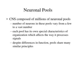
Cells of the Nervous System: Neurons • Three types of neurons: • Sensory neurons • Motor neurons • Interneurons
Neuronal structure • Soma • Dendrites • Axon • Terminal buttons • Synaptic cleft
Neuronal classifications • Multipolar neuron • Bipolar neuron (a) • Unipolar neuron (b)
Internal structure • Cell membrane • Cytoplasm • Mitochondria • Nucleus • Chromosomes • Proteins • Microtubules
Cells of the Nervous System: Glia • Glial cells support neural function
Glial Cells • Astrocytes • Arms wrap around blood vessels and neuronal structures • Isolate the synaptic cleft • Maintain chemical composition of extracellular space • Clean up following cell death
Glial cells • Oligodendrocytes (CNS) & Schwann cells (PNS) • Provide support and insulation in the form of the myelin sheath • Myelin • Nodes of Ranvier
Glial cells • Microglia • Smallest glial cells • Brain’s immune cells
Blood-Brain Barrier • Composed of tightly-packed cells of the cerebral blood vessels. • Regulates chemicals in the CNS • Protects the brain from toxins • Semipermeable
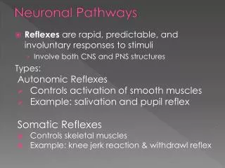
The withdrawal reflex: an example of neuronal communication • Sensory neuron detects • Message is sent • Neurotransmitter is released • Interneuron • Motor neuron sends a message
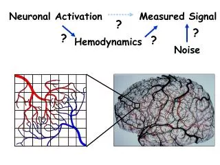
Communication within a neuron • Based on changes in the membrane potential of the neuron. • Neurons have two basic potentials
Resting membrane potential • The inside of a neuron is negatively charged relative to the outside • Due to concentrations of positively and negatively charged ions in the brain • Outside the cell • Inside the cell
Resting membrane potential • A concentration gradient pulls the sodium, potassium and chloride ions toward the membrane; electrostatic forces prevent them from crossing it. • The balance between potassium and sodium ions in and out of the neuron is maintained
Depolarization Resting Membrane Potential Hyperpolarization Membrane potential • The membrane potential can change: • Depolarization • Hyperpolarization
Action potential • A hyperpolarization _________ communication within a cell. • A depolarization _________ the cell, and _________ the chances of communication within the cell. • Threshold of excitation
Action potential • A massive, momentary reversal of the membrane potential. • Carried down the axon from the cell body to the terminal buttons. • Results in the release of a chemical message into the synapse.
Action potential • Chemical messages from other neurons affect the neuron’s charge. • Excitatory Post-Synaptic Potentials (EPSPs) • Inhibitory Post-Synaptic Potentials (IPSPs) • When the cell is depolarized to -65mV, an action potential begins.
Steps of the action potential • Ion channels in the membrane rapidly open and Na+ enters the cell (-65mV +40mV) • As Na+ rushes in, K+ is forced out of the cell. • As the action potential peaks, Na+ channels close, and no more Na+ enters the cell. • K+ is forced out of the cell, which decreases the charge inside the cell and K+ channels close. • K+ ions trapped outside of the cell result in a temporary hyperpolarized membrane potential. • Ion channels reset and the Na+/K+ pump returns the ions to the normal gradients.
All-or-None law • An action potential either occurs or it doesn’t. • Magnitude is the same. • Does not diminish in strength.
Rate law • The strength of a response depends on the firing rate of the cell. • More action potentials/second = strong response, fewer = weak response.
Action potential conduction • Action potentials depend on sodium influx from the extracellular fluid. • Nodes of Ranvier. • Saltatory conduction
Communication between neurons • Within-neuron communication: electrical signal • Between-neuron communication: chemical signal • Synaptic transmission
Synaptic structure • Presynaptic membrane • Terminal button • Vesicles • Transporter molecules • Synaptic cleft • Postsynaptic membrane • On the dendrite, soma or axon • Receptors
Neurotransmitter binding • Binding sites • Ligands • Molecule that fits into a specific binding site • Endogenous ligands • Exogenous ligands
Synaptic firing • Initiated by an action potential in the cell • Neurotransmitter (NT) binds to the receptor • Prompting specific ion channels to open
Ion Channel Neurotransmitter Gate Joins the ion channel Receptor G-protein Enzyme Second messenger Types of receptors • Ionotropic receptor • Metabotropic receptor
Synaptic firing • Postsynaptic potentials are produced by the flow of ions in and out of the cell. • Each NT produces a specific postsynaptic potential • Excitatory NTs • Inhibitory NTs
Synaptic firing • Neural integration is the summation of all postsynaptic potentials. • Determines the response to PSPs.
Synaptic firing • Remember – Each neuron has synaptic connections with hundreds of other neurons, and must summate all incoming PSPs thousands of times each second!
Synaptic firing • Removal of NT from the synapse terminates PSPs • Reuptake • Enzymatic deactivation
Autoreceptors • Found on the presynaptic membrane
Types of synapses • Axodendritic • Axosomatic • Axoaxonic