Download
1 / 62
640 likes | 1.01k Views
Chapter 7 AXIAL SKELETON. Skeletal System GROSS ANATOMY. OBJECTIVES. Distinguish between the axial and appendicular skeletons and name the bones of each. Explain the types of vertebra, the curvature of the vertebra, and number them. Explain the types of ribs and number them.

E N D
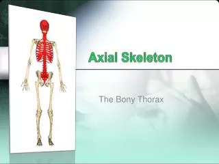
Chapter 7AXIAL SKELETON Skeletal System GROSS ANATOMY
OBJECTIVES • Distinguish between the axial and appendicular skeletons and name the bones of each. • Explain the types of vertebra, the curvature of the vertebra, and number them. • Explain the types of ribs and number them. • Explain the differences between the male and female skeleton’s pelvic cavity. • Are their any additional variations in other locations in the body?
WAY COOL WEBSITE • Body Online • http://www.innerbody.com/image/skelfov.html
Anatomical Terms used for Bone Featurespage 1 • Terms • Body: main part • Head: enlarged end • Neck: constriction between head and body • Marginorborder: edge • Angle: bend • Ramus: branch off body • Condyle: smooth rounded articular surface • Facet: small flattened articular surface • Projections • Process: prominent projection • Tubercle: small rounded bump • Tuberosity: knob • Trochanter: tuberosities on proximal femur • Epicondyle: near or above condyle
Anatomical Terms used for Bone Featurespage 2 • Ridges • Lineorlinea: low ridge • Crestorcrista: prominent ridge • Spine: very high ridge • Openings • Foramen: hole • Canalormeatus: tunnel • Fissure: cleft • Sinusorlabyrinth: cavity • Depressions • Fossa: general term for a depression • Notch: depression in bone margin • Fovea: little pit • Grooveorsulcus: deeper, narrow depression
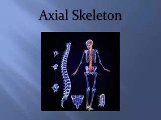
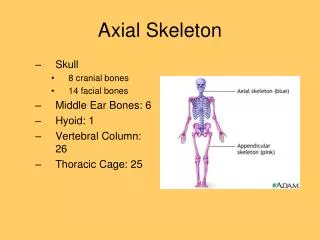
General Information • There are 206 bones of the human body. • The bones of the skeleton are divided up into • AXIAL BONES • APPENDICULAR BONES
AXIAL SKELETONgeneral bones • Skull • Auditory ossicles • Hyoid Bone • Vertebral Column • Rib Cage (thoracic cage) 80 bones
APPENDICULAR SKELETONgeneral bones • Contains: • Pectoral Girdle • Upper Limb • Pelvic Girdle • Lower Limb • Function: • Protects the Brain, Spinal Cord & Vital Organs located in the Thorax 126 Total Bones
AXIAL SKELETON SKULL
Skull (aka: Cranium) • Neurocranium (Braincase) • Viscerocranium (Face)
Neurocranium • Parietal • Temporal • Frontal • Sphenoid • Occipital • Ethmoid
Parietal Bone Coronal Suture Lamboid Suture Squamous Suture
Temporal Bone Landmarks External AcousticMeatus (ear canal) Zygomatic Process Mastoid Process Styloid Process
Temporal Bone Landmarks Carotid Canal (Carotid Foramen)
9) Internal Acoustic Meatus Temporal Bone Landmarks
Frontal Bone & Landmarks Supraorbital Margin
Sphenoid Bone & Landmarks SellaTurcica(houses pituitary) Resembles a bat or butterfly
Occipital Bone & Landmarks Opening where brain & spinal cord connect Occipital Condyle (articulation {meeting of two bones} between the skull & 1st Vertebra) Foreman Magnum
Ethmoid Bone Landmarks Middle Nasal Concha Inferior Nasal Concha
Fontanals Closes at end of 2nd year Closes during the 1st year
Viscerocranium (face)Bones of the Face • Maxilla • Zygomatic – “cheekbone” • Palatine • Lacrimal • Nasal • Inferior Nasal Concha • Mandible • Vomer
MandibularCondyle Mandible Landmarks
Maxilla Landmarks Incisive Foramen Palatine Process
Unlike other bones, the hyoid does NOT articulate with other bones. Hyoid Bone Its name is derived from the Greek word hyoeides meaning "shaped like the letter upsilon" (υ).
SINUSES • Frontal • Maxillary • Ethmoidal • Sphenoidal
Terms • Wormian Bones • also known as extra sutural bones[1] are extra bone pieces that occur within a suture in the cranium. These are irregular isolated bones which appear in addition to the usual centers of ossification of the cranium and, although unusual, are not rare.
Terms • Foramen – hole • Condyle – smooth, rounded articular surface • Articular – of or relating to joints • Process – prominent projection • Fossa – general term for a depression • Canal/Meatus – tunnel
Functions of the Vertebral Column • Supports weight • Protects the spinal cord • Allows spinal nerves to exit the spinal cord • Provides site for muscle attachment • Permits movement of head & trunk
Vertebral Columngeneral information • Consists of 26 bones called “vertebrae” • Vertebrae can be divided into 5 regions: • (C) Cervical vertebrae (7) • (T) Thoracic vertebrae (12) • (L) Lumbar vertebrae (5) • Sacral bone (1) • Coccygeal bone (1) *hint to remember the numbers think of mealtimes (7, 12, and 5)
Developing embryo have about 33-34 vertebrae that fuse: 5 sacral fuse to form 1 bone
Counting Vertebrae • The vertebrae are designated by a letter (C, T or L) with a number after the number. • The number indicates the number of the vertebrae from superior to inferior within each region.
Curvature of the Vertebral Column • There are 4 major curvatures which help accommodate our upright posture by aligning our body weight with our pelvis and lower limbs. • 2 curvatures appear during embryonic development
First Cervical Vertebrae • The first cervical (neck) vertebra is called the atlas. It supports the head. • The atlas bone is named for the Greek god Atlas who was condemned to support the earth and its heavens on his shoulders.
Second Cervical Vertebrae • The second cervical vertebra is called the axis. It is so-named because the uppermost cervical vertebra (called the atlas) rotates about the odontoid process of the second cervical vertebra. • The joint between the axis and atlas is a pivot type of joint. It allows the head turn. • The Latin word "axis" means axle or pole. • The axis bone serves as the axle about which the atlas (and the head) turn. Dens (aka: Odontoid Process)
Transverse Foramen Transverse Foramen – indicated by the BLUE ARROWS
Anatomy of Vertebrae • Body – weight bearing portion • Arch – projects posteriorly from the body • Various Processes