Download
1 / 52
670 likes | 1.59k Views
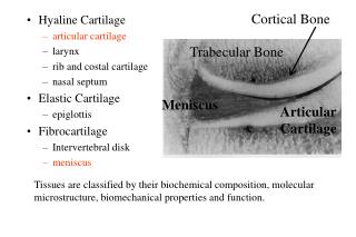
Articular Cartilage. Structure, Composition, Function. Composition. Sparse population of cells - chondrocytes Large extracellular Matrix -water -proteoglycan -collagen. Comparing Skeletal Tissue Composition. Structure. Lamina Splendens. Superficial tangential zone. Zonal Topography.

E N D
Articular Cartilage Structure, Composition, Function
Composition Sparse population of cells - chondrocytes Large extracellular Matrix -water -proteoglycan -collagen
Structure Lamina Splendens Superficial tangential zone
Zonal Topography • STZ • Parallel collagen fibrils • Flattened cells • High water • Middle Zone • Less organized, larger diameter collagen fibrils • Rounded cells • Deep Zone • Perpendicular collagen fibrils • Highest proteoglycan content • Rounded cells arranged in columns • Calcified Zone • Subchondral Bone
Proteoglycans are complex macromolecules • Protein core • Polysaccharide chains
Macromolecular Proteoglycan Structure
Nutrition & Articular Cartilage • Main source originates from vascularity in the synovium • Factors, vitamins, minerals, carbohydrates, metabolites rapidly diffuse through the synovial fluid • Diffusion through the cartilage matrix is significantly slower
Important Growth Factors • PDGF • Stimulator of mitogenesis • Only important in OA and lacerative injury • bFGF • Powerful mitogen • Works most effectively with other factors • IGF-I and II • Mitogenic and anabolic (matrix inducer) • Maintains steady proteoglycan synthesis • TGF-b • Complex constellation of actions • Alterations in signaling correlate with OA
Degradative Enzymes Important in Cartilage • Metalloproteinases • Collagenase, gelatinase, stromelysin • Depend on zinc binding • Collagenase targets triple helical collagen • Gelatinase targets individual collagen a chains • Stromelysin targets col2 and 9 and possibly aggrecan • Cathepsins/Aggrecanases • Common forms include cathepsin D and B and aggrecanase 1 and 2 (ADAMTS 4 and 5) • Exclusively targets aggrecan
Articular Cartilage:Development and Aging immature maturing adult
Most Important Biomechanical Consideration Na+, Ca2+ Donnan osmotic pressure H2O
Effects of Joint Loading and Motion • Reduced loading (immobilization) = atrophy • Continuous static compression induced lesion and chondrocyte apoptosis • Single high impact or repetitive trauma induces catabolism • Repetitive moderate loading (e.g. running) thought to be anabolic for proteoglycan • Failure of structural mechanisms induces catabolism • How loading influences chondrocyte function is unknown
Growth Plate Cartilage & Endochondral Ossification
Endochondral Bone Formation Growth plate chondrocyte differentiation • Complex interplay of intercellular • signals that co-ordinate • proliferation • hypertrophy • ossification Resting Proliferating Hypertrophic • TGF-b • BMPs • Retinoic Acid • PTHrP • Ihh • Wnts • Cytokines Bone
Type X Collagen MMP13 Alkaline Phosphatase Osteocalcin VEGF Apoptosis Type II Collagen Stages of Chondrocyte Maturation Proliferative - Prehypertrophic Terminal Maturation Undifferentiated Hypertrophic Growth Plate Chondrocytes TGF-b BMP
BMP-2 Stimulates Chondrocyte Maturation Chick Caudal Sternal Chondrocytes treated for 8 days 10 25 0 100 50 BMP-2 (ng/ml) Type X 28s rRNA Li, et al., Endocrinology 144: 2514-23, 2003
TGF-b Inhibits Chondrocyte Maturation Chick Cephalic Sternal Chondrocytes 24h 48h 72h 96h 24h 48h 72h 96h TGF-b - - - - + + + + colX 18s RNA Ionescu, et al., Exp. Cell Res. 288:198-207, 2003
P Smad 2,3 Smad4 TGF-b responsive genes TGF-b and BMP Activate Smad Pathways TGF-b receptor BMP receptor Smad 2,3 Smad 1,5 P P Smad 1,5 Smad 2,3 Smad4 Smad4 P Smad 1,5 Smad4 BMP-2 responsive genes
TGF-b Induces Nuclear Localization of Smad2 and 3 Control TGF-b BMP-2 Smad2 Smad3
What in vivo evidence is there that these signaling pathways are important in regulating maturation of chondrocytes? Smad3 deficient mice have accelerated chondrocyte maturation and OA.
How is TGF-b signaling effected in chondrocytes isolated from the neonatal sternum of wild type and Smad3-/- mice? Assessment of signaling using a TGF-b-responsive promoter/reporter
Measuring activation of TGF-b/Smad signaling induced by TGF-b 4xSBE luciferase P3TP-luc 1) Transfect 2) Treat with TGF-b 3) Measure luciferase luminescence
Activation of the SBE-Luc Reporter in Smad3-/- Chondrocytes is Completely Blocked 2000000 1800000 1600000 1400000 SBE Luciferase 1200000 1000000 800000 600000 400000 200000 0 WT KO – + – + TGF-
What is the phenotype of chondrocytes isolated from the neonatal sternum of wild type and Smad3-/- mice? Assessment of phenotypic gene expression
colX Expression is Elevated in Smad3-/- Chondrocytes 0.6 0.5 0.4 WT colX Expression 0.3 KO 0.2 0.1 0 2 Days 4 Days 8 Days colX 28S RNA
Other markers of maturation are up-regulated in Smad3-/- Chondrocytes 3.5 3 2.5 Relative Expression by RT-PCR (compared to b-actin control) 2 WT KO 1.5 1 0.5 0 AP MMP-9 VEGF-A MMP-13 Osteocalcin
What other in vivo evidence is there that the BMP/TGF-b signaling pathways are important in regulating maturation of chondrocytes?
BMP signaling induction of the transcription factor Runx2 is critical for terminal hypertrophy of chondrocytes and skeletal mineralization Inactivating mutations of the Runx2 gene are linked to the development of cleidocranial dysplasia WT Runx2 KO
Osteoarthritis Proliferative - Prehypertrophic Terminal Maturation Undifferentiated Hypertrophic Growth Plate Chondrocytes TGF-b BMP Articular Chondrocytes osteoarthritis Sox9 col2 aggrecan/ proteoglycans Ihh colx alk phos BMP-6 MMP9, 13 VEGF OC apoptosis matrix calcification
Chondrocytes Express Hypertrophic Markers During Osteoarthritis • During OA, articular chondrocytes exhibit: • - Increased proliferation (cloning) • Expression of MMPs, colX, BMP-6 and other hypertrophic markers • - Terminal hypertrophy and apoptosis BMP-6 Immunostain
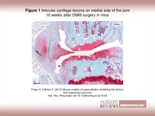
Cloning, Fibrillation and Ulceration OA normal
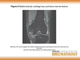
Definition and Pathology • Progressive loss of articular cartilage without a major inflammatory component • Focal fibrillation and ulceration • Cartilage swelling due to ‘loosening’ of the collagen matrix leading to increased Donnan osmosis • Cartilage loss and destruction • Subchondral sclerosis • Cyst and osteophyte formation
Etiology • Aging • Alterations in matrix • Alterations in cell activity/function • Alterations in cell mediators • Altered joint mechanics • Immune responses