Download
1 / 33
380 likes | 1.1k Views
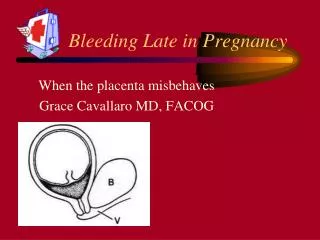
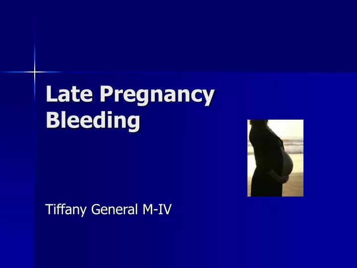
Late Pregnancy Bleeding. Tiffany General M-IV. Overview . Discuss two of the most common causes of late pregnancy bleeding Presentation Classification Risk factors Presentation Diagnosis Imaging studies Management Conclusion. Case 1.

E N D
Late Pregnancy Bleeding Tiffany General M-IV
Overview • Discuss two of the most common causes of late pregnancy bleeding • Presentation • Classification • Risk factors • Presentation • Diagnosis • Imaging studies • Management • Conclusion
Case 1 • A 40-year-old woman, 29 weeks pregnant, presented to the emergency room with painless vaginal bleeding. This is the patient's fourth pregnancy (G4 P3), and three prior births were by cesarean section. What’s the Diagnosis??
Placenta Previa • Placental plantation that overlies or is within 2 cm (0.8 in) of the internal cervical os • Classification • Complete: Placenta completely covers the os • Partial: Placenta partially covers the os • Marginal: Placenta edge lies within 2 cm of the os • Low lying: Placenta edge lies 2 to 3.5 cm from the os • Normal – positioned away from cervix (posterior position)
Placenta Previa Partial Low lying Complete
Incidence of Previa • 1 in 200-250 live births • Complete 20-45%, partial 30%, marginal 25-50% • U/S at 18 weeks shows 12-25% incidence of low lying placenta • Most of these (~90%) resolve by term • “placental migration” – placenta grows towards best blood supply located in upper uterine segment away from cervix
Risk Factors of Previa • Chronic hypertension • Multiparity • Multiple Gestations • Increased maternal age • Previous cesarean delivery • Tobacco use • Uterine curettage
Presentation • Sudden, painless, and profuse vaginal bleeding in pregnancy during the second/third trimester (usually after 28 weeks) • Thought to occur from placental detachment due to thinning of lower uterine segment in preparation for labor and/or during labor • Often bright red blood • First bleed • Usually not significant to cause homodynamic instability or threaten fetus • Rarely maternal death
Diagnosis • Any pregnant woman who presents with significant vaginal bleeding needs evaluation • History and Physical • Never do digital exam without knowing placental placement! • Could cause life-threatening hemorrhage • Most common imaging study used for diagnosis is ultrasound (ultrasonography) • Most useful and inexpensive • Transvaginal provides almost 100% accuracy in identification, transabdominal 95% • Sterile speculum exam can be done to evaluate for ruptured membrane
Diagnosis • Ultrasound - imaging method that uses high-frequency sound waves to produce precise images of structures • Cyclic sound pressure that is greater then the upper limit of human hearing (~20 kilohertz) • Images shown as thin flat sections of the body • Advancements include 3-D images and 4-D images with motion • Does not use ionizing radiation (x-ray) • Main imaging study used throughout pregnancy • Little risk to mother or fetus • Doppler ultrasound is a special ultrasound technique that evaluates blood as it flows through a vessel
Diagnosis Can you see the placenta previa? placenta cervix
Diagnosis More examples…
Diagnosis And more…
Diagnosis • MRI – not often used, but can be of benefit if placenta accreta is also suspected • Large trials concerning safety and efficacy have not been done
Diagnosis T2
Management • Women with significant vaginal bleeding are hospitalized for evaluation • Bleeding will usually resolve, but may return with the onset of labor • maternal/fetal status unstable → delivery by c-section • Maternal/fetal status stable → expected management • Cesarean is the method of delivery for placenta previa • This decision is not made until after 36 weeks because often the placenta will migrate
Case 2 30-year-old woman, gravida 3, para 2 presented at 32 weeks' gestation with a history of dull aching pain in the abdomen radiating to her back. There was no history suggestive of tightening. There was no history of vaginal bleeding. Fetal movements were normal. Patient has a history of cocaine abuse. What’s the diagnosis??
Placental Abruption (Abruptio placentae ) • Placental Abruption is the separation of the placenta from the uterine wall before delivery • Marginal separation • Partial separation • Complete separation with concealed hemorrhage • Classification • Class 0 is asymptomatic • Diagnosis is made retrospectively by finding an organized blood clot or a depressed area on a delivered placenta • Class 1 is mild and represents approximately 48% of cases • No vaginal bleeding to mild vaginal bleeding • Class 2 is moderate and represents approximately 27% of cases • No vaginal bleeding to moderate vaginal bleeding • Class 3 is severe and represents approximately 24% of cases • No vaginal bleeding to heavy vaginal bleeding
Placental Abruption Visible bleeding Concealed bleeding
Incidence of Abruption • Most common cause of late pregnancy bleeding • Occurs in 1 in 150 births • 1-3% of all births • 50% occur before 36 weeks • 80% occur before the onset of labor • Increased risk of maternal/fetal death • 10-30% neonatal mortality associated
Risk Factors of Abruption • Chronic hypertension • Multiparity • Preeclampsia • Advanced maternal age • Previous abruption • Short umbilical cord • Sudden decompression of an overdistended uterus • Thrombophilias • Tobacco, cocaine, or methamphetamine use • Trauma: blunt abdominal or sudden deceleration • Unexplained elevated maternal alpha fetoprotein level • Uterine fibroids
Presentation • Women often present with the following: • Painful vaginal bleeding • Bleeding may not be visible • Abdominal or back pain and uterine tenderness • Fetal distress • Non-reassuring fetal heart rate • Abnormal uterine contractions ( hypertonic, high frequency) • Idiopathic premature labor • Fetal death • DIC may result from the release of thromboplastin into the maternal circulation with placental separation
Diagnosis • Any pregnant woman who presents with significant vaginal bleeding needs evaluation • History and Physical • Never do digital exam without knowing placental placement! • Ultrasound will show abruption in 50% of cases • Often hard to distinguish clots on U/S • Diagnosis is based on clinical picture once other causes have been excluded • May not have definite diagnosis until clot or indentation is seen on placenta after delivery
Diagnosis Can you see the abruption? Abruption
Diagnosis More examples….
Diagnosis And more….
Diagnosis MRI – abruption with incidental previa T2 T1
Diagnosis Placental abruption after delivery
Management • If fetus is mature, homodynamic stabilization is warranted with prompt delivery • If fetus is premature, may observe with close monitoring as long as no fetal/maternal distress • Careful monitoring for uteroplacental insufficiency is essential • Must rule out coagulopathy - Check PT/PTT, platelet, fibrinogen, fibrin split products • DIC can occur as a result of abruption
Conclusion Previa vs. Abruption • Any pregnant woman who presents with vaginal bleeding must be evaluated • Never do digital exam without knowing placental placement! • Ultrasound
Reference • Fontaine P, Leeman L, Sakornbut E. Late Pregnancy Bleeding. American Family Physician 2005;75:8 • Bleeding During Pregnancy. The American College of Obstetricians and Gynecologist; http://www.acog.org/ • Blueprints Obstetric and Gynecology, 4th edition, Lippincott Williams and Wilkins • Department of Obstetrics and Gynecology, University of South Carolina (Mark Wild,MD)