Download
1 / 36
370 likes | 421 Views
Explore the diverse world of joints in the skeletal system, from fibrous to synovial joints, understanding their classification, structure, and movements. Learn about common joint disorders, life-span changes, and clinical applications.

E N D
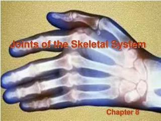
Chapter 8Joints of the Skeletal System • Articulations • Functional junctions between bones • Bind parts of skeletal system together • Make bone growth possible • Permit parts of the skeleton to change shape during childbirth • Enable body to move in response to skeletal muscle contraction
Classification of Joints • Fibrous Joints • dense connective tissues connect bones • between bones in close contact • synarthrotic • immovable • amphiarthrotic • slightly movable • diarthrotic • freely movable • Cartilaginous Joints • hyaline cartilage or fibrocartilage connect bones • Synovial Joints • most complex • allow free movement • held together by a fluid filled joint cavity
Fibrous Joints 3 Types • Syndesmosis • Suture • Gomphosis Syndesmosis • a sheet or bundle of fibrous tissue connects bones • amphiarthrotic • lies between tibia and fibula
Fibrous Joints Suture • between flat bones • synarthrotic • thin layer of connective tissue connects bones Gomphosis • cone-shaped bony process in a socket • tooth in jawbone • synarthrotic
Cartilaginous Joints 2 Types • Synchondrosis • Symphysis Synchondrosis • bands of hyaline cartilage unite bones • epiphyseal plate (temporary) • between manubrium and first rib • synarthrotic
Cartilaginous Joints Symphysis • pad of fibrocartilage between bones • pubis symphysis • joint between bodies of adjacent vertebrae • amphiarthrotic
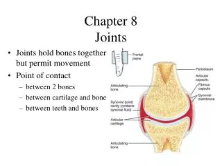
Synovial Joints • diarthrotic • joint cavity • synovial fluid • joint capsule • synovial membrane • bursae
General structure of a synovial joint • Articular cartilage covers ends of bones • Spongy bone usually beneath cartilage • Subchondral plate between bone and cartilage that absorbs shock • Joint capsule strengthened by ligaments that hold bones together • Synovial membranes secretes a clear viscous synovial fluid
Synovial fluid moistens, provides nutrients, and lubricates articular surfaces • Some areas have villi to increase surface area • Menisci divide some synovial joints into compartments • Some joints have fluid filled bursae • Bursae are usually located between the skin and underlying bony prominances
Bursae cushion and aid movement of tendons over bony parts • Bursae are named according to their locations
Types of Synovial Joints Condyloid Joint • between metacarpals and phalanges • Wide range of movement but not rotation Ball-and-Socket Joint • hip • shoulder • Widest range of motion
Types of Synovial Joints Hinge Joint • elbow • between phalanges • Moves in one plane only Gliding Joint (Plane) • between carpals • between tarsals • Permits sliding and twisting
Types of Synovial Joints Pivot Joint • between proximal ends of radius and ulna • Permits rotation Saddle Joint • between carpal and metacarpal of thumb • Permits variety of movement
Types of Joint Movements • abduction/adduction • dorsiflexion/plantarflexion • flexion/extension/hyperextension
Types of Joint Movements • rotation/circumduction • supination/pronation
Types of Joint Movements • eversion/inversion • protraction/retraction • elevation/depression
Shoulder Joint • ball-and-socket • head of humerus • glenoid cavity of scapula • loose joint capsule • bursae • ligaments prevent displacement • very wide range of movement
Elbow Joint • hinge joint • trochlea of humerus • trochlear notch of ulna • gliding joint • capitulum of humerus • head of radius • flexion and extension • many reinforcing ligaments • stable joint
Hip Joint • ball-and-socket joint • head of femur • acetabulum of coxa • heavy joint capsule • many reinforcing ligaments • less freedom of movement than shoulder joint
Knee Joint • largest joint – • most complex • medial and lateral condyles of distal end of femur • medial and lateral condyles of proximal end of tibia • femur articulates anteriorly with patella • modified hinge joint (2 condyloids & gliding) • flexion/extension/little rotation • strengthened by many ligaments and tendons • menisci separate femur and tibia • bursae
Life-Span Changes • Joint stiffness is an early sign of aging • Fibrous joints first to change; can strengthen over a lifetime • Changes in symphysis joints of vertebral column diminish flexibility and decrease height • Synovial joints lose elasticity • Disuse hampers the blood supply • Activity and exercise can keep joints functional longer
Clinical Application Joint Disorders Sprains • damage to cartilage, ligaments, or tendons associated with joints • forceful twisting of joint • Overstretching Bursitis • inflammation of a bursa • overuse of a joint
Joint Disorders • Arthritis • inflamed, swollen, painful joints If a joint is immobilized for a long period of time the articular cartilage may soften and degenerate Arthroscopy is used to examine a joint – blue box page 271
Rheumatoid Arthritis • autoimmune disorder • synovial membrane thickens forming a mass called a pannus • articular cartilage is damaged and bones fuse together (bony ankyloses • systemic disorder often affecting the joints, skin, eyes, lungs, blood vessels, and heart
Osteoarthritis • Most common type • Degenerative and usually occurs with aging • Articular cartilage softens and disintegrates slowly • Joints become painful with restricted movements
Lyme Arthritis • Bacterial infection passed in a tick bite • Causes intermittent arthritis of several joints several weeks after initial infection • Symptoms first appear as a rash, fatigue, flulike aches and pains • Treated with antibiotics • May be difficult to diagnose