Download
1 / 20
240 likes | 1.07k Views
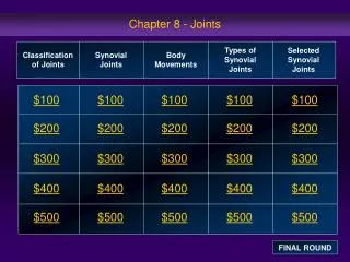
Chapter 8 Joints. Joints hold bones together but permit movement Point of contact between 2 bones between cartilage and bone between teeth and bones. Classification of Joints . Structural classification based upon: presence of space between bones (synovial cavity)

E N D
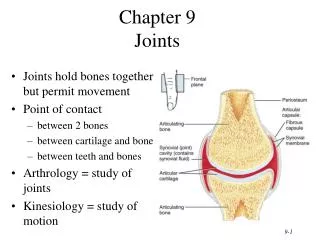
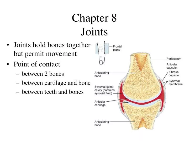
Chapter 8Joints • Joints hold bones together but permit movement • Point of contact • between 2 bones • between cartilage and bone • between teeth and bones
Classification of Joints • Structural classification based upon: • presence of space between bones (synovial cavity) • type of connective tissue holding bones together • Fibrous joints- collagen fibers • Cartilaginous joints-cartilage • Synovial joints-joint capsule & accessory ligaments • Functional classification based upon movement: • immovable = synarthrosis • slightly movable = amphiarthrosis • freely movable = diarthrosis (synovial joints)
Fibrous Joints • Lack a synovial cavity • Bones held closely together by fibrous connective tissue • Little or no movement (synarthroses or amphiarthroses) • 3 structural types • sutures • syndesmoses • gomphoses
Sutures • Thin layer of dense fibrous connective tissue unites bones of the skull • Immovable (synarthrosis) • If fuse completely in adults is synostosis
Syndesmosis • Fibrous joint • bones united by ligament • Slightly movable (amphiarthrosis) • Anterior tibiofibular joint and Interosseous membrane
Gomphosis • Ligament holds cone-shaped peg in bony socket • Immovable (synarthrosis) • Teeth in alveolar processes
Cartilaginous Joints • Lacks a synovial cavity • Allows little or no movement • Bones tightly connected by fibrocartilage or hyaline cartilage • 2 types • synchondroses • symphyses
Synchondrosis • Connecting material is hyaline cartilage • Immovable (synarthrosis) • Epiphyseal plate or joints between ribs and sternum
Symphysis • Fibrocartilage is connecting material • Slightly movable (amphiarthroses) • Intervertebral discs and pubic symphysis
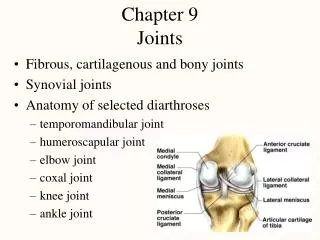
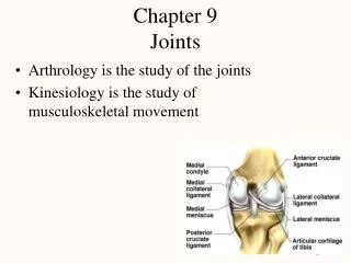
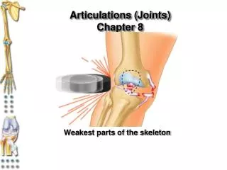
Synovial Joints • Synovial cavity separates articulating bones • Freely moveable (diarthroses) • Articular cartilage • reduces friction • absorbs shock • Articular capsule • surrounds joint • thickenings in fibrouscapsule called ligaments • Synovial membrane • inner lining of capsule • secretes synovial fluid containing hyaluronic acid slippery) • brings nutrients to articular cartilage
Example of Synovial Joint • Joint space is synovial joint cavity • Articular cartilage covering ends of bones • Articular capsule
Other Special Features • Accessory ligaments • extracapsular ligaments • outside joint capsule • intracapsular ligaments • within capsule • (A. & P. cruciate ligaments) • Articular discs or menisci • pads of fibrocartilage • attached around edges to capsule • allow 2 bones of different shape to fit tightly • increase stability of knee - torn cartilage • Bursae = saclike fluid-filled structures • skin/bone or tendon/bone or ligament/bone • knuckle cracking?
Arthroscopy & Arthroplasty • Arthroscopy = examination of joint • instrument size of pencil • remove torn knee cartilages & repair ligaments • small incision only • Arthroplasty = replacement of joints • total hip replaces acetabulum & head of femur • plastic socket & metal head • knee replacement common
Nerve and Blood Supply • Nerves to joints are branches of nerves to nearby muscles • Joint capsule and ligaments contain pain fibers and sensory receptors • Blood supply to the structures of a joint are branches from nearby structures • supply nutrients to all joint tissues except the articular cartilage which is supplied from the synovial fluid
Sprain versus Strain • Sprain • twisting of joint that stretches or tears ligaments • no dislocation of the bones • may damage nearby blood vessels, muscles or tendons • swelling & hemorrhage from blood vessels • ankle is frequently sprained • Strain • less serious injury • overstretched or partially torn muscle
Bursae and Tendon Sheaths • Bursae • fluid-filled saclike extensions of the joint capsule • reduce friction between moving structures • skin rubs over bone • tendon rubs over bone • Tendon sheaths • tubelike bursae that wrap around tendons at wrist and ankle where many tendons come together in a confined space • Bursitis • chronic inflammation of a bursa
Range of Motion in a Synovial Joint • Shape of articulating bones • Tension & strength of joint ligaments • Arrangement of muscles around joints • Apposition (coming together) of soft parts • Hormones • relaxin from placenta loosens pubic symphysis • Disuse • decreased synovial fluid, decreased flexibility of ligaments, reduced size of muscles
Rheumatoid Arthritis • Autoimmune disorder • Cartilage attacked • Inflammation, swelling & pain • Final step is fusion of joint
Osteoarthritis • Degenerative joint disease • aging, wear & tear • Noninflammatory---no swelling • only cartilage is affected not synovial membrane • Deterioration of cartilage produces bone spurs • restrict movement • Pain upon awakening--disappears with movement