Download
1 / 42
430 likes | 744 Views
Chapter 11. The Brain & Spinal Cord. Introduction. Brain & s.c. comprise the CNS Brain is protected by cranium & meninges – membranes that surround brain Consists of 3 layers: 1. dura mater 2. arachnoid mater 3. pia mater. Meninges.

E N D
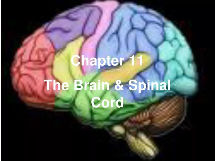
Chapter 11 The Brain & Spinal Cord
Introduction • Brain & s.c. comprise the CNS • Brain is protected by cranium & meninges – membranes that surround brain • Consists of 3 layers: 1. dura mater 2. arachnoid mater 3. pia mater
Meninges 1. Dura mater – outermost; tough, fibrous; attached to inside of cranium; contains many b.v. & nerves Arachnoid mater – thin & weblike; no b.v. or nerves SUBARACHNOID SPACE – contains cerebrospinal fluid (CSF) 3. Pia mater – delicate; w/b.v. & nerves; attached directly to brain & follows contours
Importance of Meninges • dural sinus – space formed when d.m. splits into 2 layers • subdural hematoma – fluid & blood collects under d.m. from trauma • Meningitis – inflammation of arachnoid or pia mater from bacteria or virus
Partitions of Dura mater • Falx cerebelli – b/t rt. & lt. cerebellar hemispheres 2. Falx cerebri – b/t rt. & lt. cerebral hemispheres • Tentorium cerebelli – b/t cerebrum & cerebellum
Protection of Spinal Cord • S.C. protected by bony vertebrae & same 3 meninges • Epidural space – b/t vertebrae & d.m.; contains b.v. and connective tissue for protection
The Spinal Cord • Consists of 31 segments • Each gives rise to a spinal nerve • Provides 2-way communi- cation b/t brain & body • 2 main functions: 1. center for reflexes 2. conducts impulses to & from brain
The Spinal Cord • Beginning pt. – foramen magnum • Ending pt. – conus medullaris (narrow pt. b/t L1-L2) • Cauda equina – cord of connective tissue (a.k.a. filium terminale)
Cross Section – Spinal Cord • Gray matter – horns • White matter – funiculi • 2 grooves divide s.c. into rt. & lt. halves: posterior median sulcus anterior median fissure
Cross Section - S.C. • Central canal – contains CSF; continuous w/spaces in brain • Gray commissure – connects “wings” of “butterfly”
Nerve Tracts • White matter in s.c. consists of fibers called nerve tracts; provide 2-way communication b/t brain & s.c.; • 2 types: 1. ascending – sensory fibers carry impulses to brain *In the medulla, fibers cross over
Nerve Tracts 2. descending – motor fibers carry impulses to muscles * In the medulla, fibers cross over
Reflexes • S.C.- center for reflexes (automatic, subconscious responses) • Reflexes control many involuntary actions (HR, resp.rate, swallowing, sneezing, etc.) • Pathway that neurons follow in a reflex - reflexarc. • One of the simplest – patellar reflex (helps maintain an upright position) • Involves only 2 neurons, sensory & motor (no interneuron)
Parts of a Reflex Arc • Most reflexes include 5 structures: 1. receptor 2. sensory n. 3. interneuron 4. motor neuron 5. effector • Other examples: withdrawal reflex (occurs when a person touches something painful) plantar reflex, Babinski reflex (abnormal in adults), biceps, triceps & ankle jerk reflexes
Ventricles of Brain • Ventricles - Interconnected cavities in brain - continuous w/central canal & subarachnoid space of s.c. ; filled w/CSF • 4 ventricles: 1st (left hemisphere) 2nd (rt. hemisphere) 3rd (midline of brain) 4th (in brainstem)
Pathway of CSF Circulation 1. Most CSF produced in lat. ventr. by choroid plexuses 2. Interventricular foramina – openings; allow CSF to flow from lat. to 3rd ventr. 3. 3rd ventricle 4. Cerebral aqueduct – narrow canal; connects 3rd to 4th ventr. 5. 4th ventricle
CSF Circulation • Flows into central canal & SA space of s.c. & back to subarachnoid space of brain 7. CSF reabsorbed by arachnoid granulations 8. Drain into blood-filled dural sinus into circ. sys. Humans secrete approx. 500ml of CSF daily. Only about 150 ml in CNS at any given time (continuously reabsorbed) CSF - clear fluid; nourishes cells of the CNS; completely surrounds brain & s.c. for protection.
Lumbar Puncture • Needle inserted into subarachnoid space of s.c. & CSF is withdrawn • Site is usually b/t L1-L2 or L3-L4 (a.k.a. spinal tap) • A manometer used to measure CSF pressure • CSF can be analyzed for viruses, bacteria, bleeding, tumors of the n.s., MS, & early-onset Alzheimers
Normal vs. Hydrocephalic Brain ←Normal Normal Brain Hydrocephaly Normal intracranial Excessive accumulation pressure 7-15 mm Hgof CSF causes ventricles in brain to dilate; infant’s skull expands & incr. in circumference (bulging fontanels possible)
Treatment of Hydrocephaly • Shunt placed in brain to regulate pressure & reabsorb CSF into subarachnoid space
The Human Brain • 5 Major Areas: 1. Cerebrum 2. Basal ganglia 3. Diencephalon 4. Brain stem 5. Cerebellum
Cerebrum • Largest part of brain • Consists of 2 halves (hemi- spheres) • Connected by corpus callosum (collection of nerve fibers) • Convolutions – raised ridges • Sulci – shallow grooves • Fissures – 2 deep grooves 1. Longitudinal – divides brain into rt. & left halves
Cerebrum • Transverse – separates cere- brum from cere- bellum • Cerebral cortex – thin, outer gray matter; contains cell bodies • White matter – under gray; makes up most of the cerebrum
Functions of Cerebrum • 3 basic functions: 1. Motor area – sends impulses to muscles 2. Sensory area – interpret impulses from sensory receptors 3. Association area – not primarily motor or sensory; interprets, analyzes, reasons, memory, problem solving, etc.
Lobes of the Brain • Sulci divide each cerebral hemisphere into 5 functional areas called lobes (named for skull bones). • 5th lobe - insula (not shown) located deep w/in lateral sulcus & covered by parts of frontal, parietal & temporal lobes
Lobes of the Brain • Frontal • Association areas – problem solving, planning, analyzing, etc. • Motor areas – (ant. to central sulcus) – control of voluntary muscles • Broca’s area – ant. to motor cortex & in left hemi.; coordinates muscles of speech
Lobes of the Brain 2. Parietal – • Somato- sensory area – cutaneous & other senses • Association area – under- standing speech & using words
Lobes of the Brain 3. Occipital – visual area 4. Temporal – auditory area & auditory memories • Wernicke’s area – in left temporal lobe; controls analysis of spoken language 5. Insula – deep w/in lateral sulcus & includes parts of frontal, parietal & temporal lobes; associated w/emotions
Basal Ganglia • Also called basal nuclei • Consist of gray matter deep within the cere- bral hemispheres • Relay info from cortex to brainstem & s.c. • Produce the ntm dopamine that inhibits motor functions (decr. levels assoc. w/Parkinson’s disease)
Diencephalon • Includes 2 regions: • Thalamus – receives all sensory info & channels it to correct region on cerebral cortex for interpret- ation • Hypothalamus – maintains homeo- stasis (i.e. heart rate, b.p., temp., hunger, sleep & wake cycles, growth)
Limbic System • Also located in the diencephalon is the limbic system • This area controls emotions & is also assoc.w/memory
Pineal & Pituitary Glands • Also located in diencephalon • Pineal gland – secretes melatonin in absence of light • Controls sleep & wake cycles • Pituitary gland – regulates growth & reproductive cycles under direction of hypothalamus
Brainstem • Connects brain to s.c. • Includes 3 regions: 1. midbrain 2. pons 3. medulla
Midbrain • 1st, short section of brainstem • Relays info. from lower parts of b.s. & s.c. to higher brain • Contains corpora quadrigemina – structure that allows movement of eyes & head at same time
Pons • Rounded bulge on underneath side of b.s. • Sends impulses to & from medulla & cerebellum
Medulla Oblongata • Enlarged contin- uation of s.c. • All nerve tracts pass thru here & many cross over • Acts as relay center b/t s.c. & cerebral cortex
Medulla • Contains 3 centers: 1. Cardiac center – area that controls heart rate 2. Vasomotor center – constricts or dilates b.v. 3. Respiratory center – regulates rate & depth of breathing • Nonvital centers – coughing, sneezing, swallowing, vomiting also located in medulla
Reticular Formation • Nerve fibers scattered throughout the b.s. • When sensory impulses reach the r.f., it responds by activating the cerebral cortex into wakefulness • The cerebral cortex can also activate the r.f. (intense cere- bral activity keeps a person awake) • If the r.f. is destroyed, a person remains in a comatose state
Reticular Formation • The r.f. filters incoming sensory info & decides what is important • Decreased activity in the r.f. results in sleep • Types of Sleep: 1. Slow-wave (non-REM)- restful, dreamless; reduced b.p. & resp. rate; lasts from 70-90 min. & alternates w/REM sleep
Sleep • REM sleep (rapid eye movement) – “paradoxical sleep”; dream sleep; lasts 5-15 min.; heart & resp. rate irregular; so important that if a person lacks it one night, it is made up for the next night
Cerebellum • Composed mostly of white matter • A treelike pattern is visible called the arbor vitae • Integrates info about body position • Coordinates skeletal muscle activity • Maintains posture & equilibrium