Download
1 / 37
470 likes | 1.12k Views
Sensory Physiology. Structure and function of receptors and effectors. How to classify sensory receptors? A. Energy transduced. Chemoreceptors Taste buds, olfactory receptors, aortic and carotid bodies Photoreceptors Rods and cones in retina Thermoreceptors Heat and cold Mechanoreceptors

E N D
Sensory Physiology Structure and function of receptors and effectors
How to classify sensory receptors?A. Energy transduced • Chemoreceptors • Taste buds, olfactory receptors, aortic and carotid bodies • Photoreceptors • Rods and cones in retina • Thermoreceptors • Heat and cold • Mechanoreceptors • Touch and pressure
B. By the type of stimulus Sensory neurons transmit specific signals; require “adequate stimulus” to do so
C. Type of sensory information delivered • Proprioreceptors • Within muscles; at joints (position and movement) • Cutaneous receptors • In skin:touch and pressure; heat and cold; pain • Special sensory organs • Eyes, ears, olfactory, taste buds • Extero- and interoreceptors • External and internal stimuli • Lots of overlap!
Sensory adaptation • Phasic (fast-adapting) and tonic (slow-adapting) receptors • Note different firing patterns • Stimulation pattern resembles EPSPs
What is a somatesthetic sensation? • From cutaneous and proprioreceptors • Myelinated sensory neurons extend all the way to medulla oblongata; crossing over • Always extends to postcentral gyrus • Referred pains along same pathway
Modulating cutaneous sensations • Receptive field: density of receptors • Some body areas • Lateral inhibition helps “pinpoint” sensation or increase acuity • Applies to other senses in addition to touch
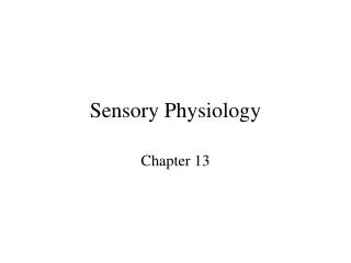
Chemoreceptors and dissolved molecules Taste and smell Gustation and olfaction
Taste buds • Have neuron-like features • Located in epithelial papillae of tongue • Cranial nerves VII and IX transmit information through thalamus to various parts of cerebral cortex
Categories of taste • All types of taste cells are found in taste buds • Each activates a specific sensory neuron • Sensory mechanism include ion channels and G-protein-linked receptors (gustducins)
Olfaction: the sense of smell • Olfactory neurons are specific • Receptors are G-protein-coupled, may be several linked to a single receptor • Olfactory information sent from olfactory bulb; project to prefrontal cortex, medial temporal lobe, hippocampus, amygdala
Vestibular apparatus and equilibrium • Otolith organs • Utricle and saccule • Semicircular canal • Membranous labyrinth encloses sensory structures • Depolarization depends on movement of K+ ions in endolymph • Bony labyrinth encloses all
The otolith organs and equilibrium • “Otoliths” are calcium carbonate crystals • Utricle sensitive to horizontal acceleration • Saccule sensitive to vertical acceleration
The middle ear conducts sound- and the stapedius muscle protects it
Arrangement of the cochlea • Cochlear duct is in scala media • Scala vestibuli and scala tympani contain perilymph • Basilar membrane and vestibular membrane bound the cochlear duct • Basilar membrane important for pitch discrimination
The organ of Corti • Stimulated by displacement of basilar membrane • Inner and outer hair cells • Tectorial membrane • More bending of stereocilia= louder sounds • Outer hair cells act as amplifiers
How do we hear sound? Cochlea analyzes pitch Neural pathways to auditory cortex
Hearing impairments • Conduction deafness • Sound waves can’t move to tympanic membrane • Middle ear damage • Affects all sound frequencies • Hearing aids • Sensorineural deafness • Damage to sensory hair cells or vestibulocochlear nerve • Cochlear implants
Accommodation contributes to visual acuity • Ciliary muscle controls shape of lens • When ciliary muscle relaxes, lens is taut and flat (distance vision) • When muscle contracts, lens becomes more round (near objects) • Loss of accommodation = presbyopia
Visual acuity • Myopia- eyeball is too long • Hyperopia- eyeball is too short • Astigmatism- curvature of lens and/or cornea is distorted
Structure and function of the retina • Rods and cones • Extension of the brain, so light passes through neurons to reach the photoreceptors • Pigment epithelium is continuously recycled • Pigments are activated by light • Rods contain rhodopsin • Rhodopsin levels increase in the dark
Light affects electrical activity of retinal cells • Inhibitory neurotransmitter is released in dark • Light prevents it • Ganglion cells perceive light if they are activated
Cones, color vision, and visual acuity • Humans are trichromats • Input passes through lateral geniculate nuclei in thalamus
Fovea centralis • Visual sensitivity is the greatest • Sensitivity to light is the lowest
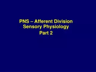
Eye movements also contribute to visual acuity • Keep image focused on fovea • Extrinsic: • Saccadic eye movements (rapid) • Smooth pursuit track moving objects • Vergence movements give depth • Fixational movements on stationary objects • Instrinsic: • Tectal (pupillary reflexes; circadian adaptation)
Where is visual information processed? • Receptive fields: ganglia receive input from one or more photoreceptors • Lateral geniculate nuclei in thalamus • Visual association areas in occipital lobe
Summary • Sensory receptors categorized on basis of structure, the stimulus they transduce, or the nature of the response. • Sensory information is carried to a specified part of the brain, by a specialized pathway, and perhaps by a specialized sensory organ • Somatesthetic- cutaneous and proprioreceptors • Taste- taste buds • Olfaction-olfactory epithelium • Equilibrium- vestibular apparatus • Hearing-the ears • Vision- the eyes