Download
1 / 43
430 likes | 473 Views
Explore the intricate workings of equilibrium, vision, and the eye's structure, from the mechanoreceptors in the vestibular apparatus to the sensory layers of the retina. Learn about static and dynamic equilibrium, maculae, cones and rods, lens accommodation, and eye chamber fluids.

E N D
Sensory Physiology equilibrium & vision
Equilibrium: Mechanoreceptor • Body balance • Body position • Body movement • Propioceptors • Vision • Vestibular apparatus
Organs of Equilibrium • Receptor cells are in two structures • Vestibule • Semicircular canals Figure 8.16a, b
Organs of Equilibrium • Equilibrium has two functional parts • Static equilibrium – sense of gravity at rest • Dynamic equilibrium – angular and rotary head movements Figure 8.16a, b
Static Equilibrium - Rest • Maculae – receptors in the vestibule • Report on the position of the head • Send information via the vestibular nerve • Anatomy of the maculae • Hair cells are embedded in the otolithic membrane • Otoliths (tiny stones) float in a gel around the hair cells • Movements cause otoliths to bend the hair cells
Function of Maculae Figure 8.15
Dynamic Equilibrium - Movement • receptors in the semicircular canals • Tuft of hair cells • Cupula (gelatinous cap) covers the hair cells Figure 8.16c
Dynamic Equilibrium • Action of angular head movements • The cupula stimulates the hair cells • An impulse is sent via the vestibular nerve to the cerebellum Figure 8.16c
Equilibrium: Mechanoreceptor • Integration • Medulla • Cerebellum • Thalamus • Cortex Figure 10-26: Central nervous system pathways for equilibrium
Equilibrium: Vestibular Apparatus • Otolith organs • Gravity • Calcite crystals • Hair cells • Semicircular canals • Fluid moves • Cristae • Cupula • Hair cells
Equilibrium: Vestibular Apparatus Figure 10-23a, b: ANATOMY SUMMARY: Vestibular Apparatus
Equilibrium: Vestibular Apparatus Figure 10-23c, d: ANATOMY SUMMARY: Vestibular Apparatus
The Eye and Vision • 70 percent of all sensory receptors are in the eyes, only see 1/6th of eye • Each eye has over a million nerve fibers • Protection for the eye • Most of the eye is enclosed in a bony orbit • A cushion of fat surrounds most of the eye
Accessory Structures of the Eye • Eyelids • Eyelashes Figure 8.1b
Accessory Structures of the Eye • Eyelashes =have modified sebacious glands produce an oily secretion to lubricate the eye Figure 8.1b
Accessory Structures of the Eye • Conjunctiva • Membrane that lines the eyelids • Connects to the surface of the eye • Secretes mucus to lubricate the eye
Accessory Structures of the Eye • Lacrimal gland • Tears: antibodies, lysozymes, stress? Figure 8.1a
Extrinsic Eye Muscles • Muscles attach to the outer surface of the eye • Produce eye movements Figure 8.2
Structure of the Eye • The wall is composed of three layers • Sclera&Cornea fibrous outside layer • Choroid – middle layer • Sensory (retina) inside layer Figure 8.3a
The Fibrous layer • Sclera • White connective tissue layer • Seen anteriorly as the “white of the eye” • Cornea • Transparent, central anterior portion • Allows for light to pass through • Repairs itself easily • The only human tissue that can be transplanted without fear of rejection
Choroid Layer • Blood-rich nutritive layer • Pigment prevents light from scattering • Modified interiorly into two structures • Cilliary body – smooth muscle • Iris • Pigmented layer that gives eye color • Pupil – rounded opening in the iris
Sensory layer (Retina) • Contains receptor cells (photoreceptors) • Rods • Cones • Signals pass from photoreceptors and leave the retina toward the brain through the optic nerve
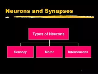
Neurons of the Retina Slide 8.11
Neurons of the Retina and Vision • Rods • Most are found towards the edges of the retina • Allow dim light vision and peripheral vision • Perception is all in gray tones
Neurons of the Retina and Vision • Cones – 3 types detect different colors • Most dense at the center of the retina • Fovea centralis – area of the retina with only cones • Lack of one type = color blindness • No photoreceptor cells are at the optic disk, or blind spot
Lens • Biconvex crystal-like structure • Held in place by a suspensory ligament attached to the ciliary body Figure 8.3a
Internal Eye Chamber Fluids • Aqueous humor in Anterior Segment • Watery fluid found in chamber between the lens and cornea • Similar to blood plasma • Helps maintain intraocular pressure • Provides nutrients for the lens and cornea • Reabsorbed into venous blood • Blocked drainage = glaucoma
Internal Eye Chamber Fluids • Vitreous humor in Posterior Segment • Gel-like substance behind the lens • Keeps the eye from collapsing • Lasts a lifetime and is not replaced
Lens Accommodation • Light must be focused to a point on the retina for optimal vision • The eye is set for distance vision (over 20 ft away) • The lens must change shape to focus for closer objects Figure 8.9
Correcting the Eye • Nearsightedness = myopia • Focus of light in front of retina • Eyeball too long or lens too thick • Distant objects are blurry • Farsightedness = hyperopia • Focus of light beyond the retina • Short eyeball or lens too thin • Near objects are blurry.
Astigmatism • Unequal curvatures in cornea & lens
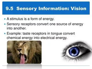
Vision: Photoreceptors • Reflected light translated into mental image • Pupil limits light, lens focuses light • Retinal rods and cones are photoreceptors Figure 10-36: Photoreceptors in the fovea
Photoreception and Local Integration • Rods – night vision • Cones – color & details • Bipolar & ganglion cells converge, integrate APs
Photoreception and Local Integration Figure 10-35: ANATOMY SUMMARY: The Retina
Retina: More Detail • Rod cells: monochromatic • Cone cells: red, green, & blue • Discs: visual pigments • Pigmented epithelium • Melanin granules • Prevents reflection
Retina: More Detail Figure 10-38: Photoreceptors: rods and cones
Phototransduction • Photons "bleach" opsin, retinal released, cascade, Na+ channel closes, K+ opens , hyperpolarization • Reduces NT release
Phototransduction Figure 10-40: Phototransduction in rods
Vision: Integration of Signals to Perception • Bipolar • Ganglion • Movement • Color • Optic nerve • Optic chiasm • Optic tract • Thalamus • Visual cortex Figure 10-29b, c: Neural pathways for vision and the papillary reflex
Summary • Sensory pathway: receptor, sensory neuron(s) & CNS • Somatic senses: touch, temperature, pain & proprioception communicate body information to CNS • Special senses: taste, smell, hearing, equilibrium, & vision • Outside conditions for CNS integration into perception • Receptors transduce mechanical, chemical or photon energy into GPs then to APs